Letter
August 2025, Volume 38, nº4
5 min
Conjunctival and cutaneous Kaposi's Sarcoma as the first manifestation of HIV infection
Raúl Parra-Fariñas1 , Carlota Insua2, Lucía Buznego3, Paula Runza1, Carlos Armiñanzas1,4, Manuel Gutierrez-Cuadra1,4, Sara Marcos-González5, Francisco Arnaiz de las Revillas1,4
1Infectious Diseases Service, Hospital Universitario Marqués de Valdecilla-IDIVAL, University of Cantabria, Santander, España
2Internal Medicine Service, Hospital Universitario Marqués de Valdecilla, Santander, España
3Oncology Service, Hospital Universitario Marqués de Valdecilla-IDIVAL, Santander, España
4Centro de Investigación Biomédica en Red de Enfermedades Infecciosas (CIBERINFEC), Instituto de Salud Carlos III, Madrid, España 5Pathology Service, Hospital Universitario Marqués de Valdecilla, Santander, España
http://www.doi.org/10.37201/req/013.2025
Receipt Date:
24/01/2025
Acceptance Date:
25/04/2025
Publication Date:
26/05/2025
How to cite
Parra-Fariñas R, Insua C, Buznego L, Runza P, Armiñanzas C, Gutierrez-Cuadra M, et al. Conjunctival and cutaneous Kaposi's Sarcoma as the first manifestation of HIV infection. Rev Esp Quimioter 2025; 38(4): 352-354. Available from: http://www.doi.org/10.37201/req/013.2025
Metrics
793
Views
50
Downloads
Share
Share
Sir,
Kaposi's sarcoma (KS) is a vascular angiosarcoma associated with chronic infection by human herpesvirus type 8 (HHV-8) [1]. It frequently arises in the setting of immunosuppression, especially in individuals with human immunodeficiency virus (HIV) infection [2-4]. However, the efficacy and widespread access to antiretroviral therapy in developed countries have made it possible to control HIV replication, leading to a significant decrease in the risk of developing KS in people living with HIV (PLHIV) over the past two decades, particularly in resource-rich settings [5].
Despite this decline in incidence, KS remains prevalent in PLHIV [6]. Nonetheless, ocular involvement by KS, as a defining manifestation of acquired immunodeficiency syndrome (AIDS), is a rare event [7].
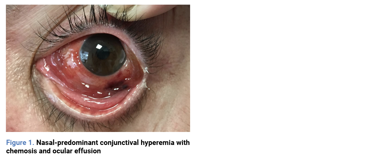
We present the case of a 45-year-old male with a history of inverse psoriasis who presented to the Emergency Department of a tertiary hospital due to pain in his left eye. Physical examination revealed conjunctival hyperemia predominantly in the nasal area, with chemosis and subconjunctival hemorrhage. He was diagnosed with conjunctivitis and discharged with topical antibiotic treatment for seven days. After completing the treatment and observing no improvement, he returned to the Emergency Department, where another cycle of topical antibiotics was prescribed, and he was referred to the Ophthalmology outpatient clinic (Figure 1).
 A blood test revealed a white blood cell count of 2.9 × 10³/µL, hemoglobin at 11.6 g/dL, and platelets at 108 × 10³/µL. One week later, he was evaluated in the Ophthalmology clinic, where he was diagnosed with scleritis and started on topical corticosteroids. A referral to Rheumatology was made due to suspected autoimmune disease.
In Rheumatology, as part of a routine workup, various serologies were requested, revealing a positive HIV-1 serology. Simultaneously, a conjunctival biopsy was performed due to the poor clinical progression, which confirmed the diagnosis of ocular KS with CD31 and HHV-8 expression. The patient was subsequently referred to the Infectious Diseases outpatient clinic, where other lesions were identified on the abdominal wall and proximal right lower limb, suggestive of KS.
The HIV viral load was 2,440,000 copies/mL, and the CD4+ T-cell count was 26 cells/µL. Antiretroviral therapy with tenofovir, emtricitabine, and raltegravir was initiated. A computed tomography scan ruled out visceral involvement at other levels. One month after starting antiretroviral therapy, the patient showed improvement in skin lesions but worsening ocular symptoms, which were interpreted as immune reconstitution inflammatory syndrome. A watchful approach was taken, and two months after initiating treatment, ocular improvement was observed. The CD4+ T-cell count had risen to 285 cells/µL, and the viral load had decreased to 11,100 copies/mL, with no cutaneous lesions present.
Six months later, the patient had no skin lesions, and on ophthalmologic examination, only a brownish discoloration persisted in the eye. Currently, after four years of follow-up, the patient has had no recurrence of either cutaneous or ocular lesions.
In PLHIV, KS generally first affects the skin, although in up to 20% of cases, early mucosal involvement is seen, predominantly in the oral cavity [1]. Historically, KS was one of the most characteristic manifestations of the AIDS epidemic, with the lesions it caused becoming one of its first visible clinical signs in the 1980s [2-4]. However, nowadays, it is rarely seen as the first manifestation in HIV-positive patients [6].
In our case, ocular KS was the first manifestation of HIV infection, and its initial diagnosis was incidental. This is because ocular involvement is a very rare presentation, requiring a high index of suspicion. In this case, the cutaneous lesions were not diagnosed as the patient associated them with his previously diagnosed psoriasis.
Although ocular KS has been described in HIV patients in earlier periods [8, 9], when antiretroviral treatments were less effective, we consider it noteworthy that, currently, in a patient with lymphopenia and conjunctival lesions unresponsive to initial ophthalmologic treatment, HIV infection should be considered in the differential diagnosis to avoid delays in diagnosis and initiation of antiretroviral therapy.
Funding
None to declare.
Conflicts of interest
The authors declare no conflicts of interest.
Authors contributions
Conceptualization, R.P.F. and F.A.R.; methodology, R.P.F. and F.A.R.; software, R.P.F. and C.I., validation, F.R.A.; writing—original draft preparation, R.P.F., and C.I.; writing—review and editing, L.B., P.R., C.A., M.G.C., S.M.G and F.R.A.; supervision, F.R.A. All authors have read and agreed to the published version of the manuscript.
A blood test revealed a white blood cell count of 2.9 × 10³/µL, hemoglobin at 11.6 g/dL, and platelets at 108 × 10³/µL. One week later, he was evaluated in the Ophthalmology clinic, where he was diagnosed with scleritis and started on topical corticosteroids. A referral to Rheumatology was made due to suspected autoimmune disease.
In Rheumatology, as part of a routine workup, various serologies were requested, revealing a positive HIV-1 serology. Simultaneously, a conjunctival biopsy was performed due to the poor clinical progression, which confirmed the diagnosis of ocular KS with CD31 and HHV-8 expression. The patient was subsequently referred to the Infectious Diseases outpatient clinic, where other lesions were identified on the abdominal wall and proximal right lower limb, suggestive of KS.
The HIV viral load was 2,440,000 copies/mL, and the CD4+ T-cell count was 26 cells/µL. Antiretroviral therapy with tenofovir, emtricitabine, and raltegravir was initiated. A computed tomography scan ruled out visceral involvement at other levels. One month after starting antiretroviral therapy, the patient showed improvement in skin lesions but worsening ocular symptoms, which were interpreted as immune reconstitution inflammatory syndrome. A watchful approach was taken, and two months after initiating treatment, ocular improvement was observed. The CD4+ T-cell count had risen to 285 cells/µL, and the viral load had decreased to 11,100 copies/mL, with no cutaneous lesions present.
Six months later, the patient had no skin lesions, and on ophthalmologic examination, only a brownish discoloration persisted in the eye. Currently, after four years of follow-up, the patient has had no recurrence of either cutaneous or ocular lesions.
In PLHIV, KS generally first affects the skin, although in up to 20% of cases, early mucosal involvement is seen, predominantly in the oral cavity [1]. Historically, KS was one of the most characteristic manifestations of the AIDS epidemic, with the lesions it caused becoming one of its first visible clinical signs in the 1980s [2-4]. However, nowadays, it is rarely seen as the first manifestation in HIV-positive patients [6].
In our case, ocular KS was the first manifestation of HIV infection, and its initial diagnosis was incidental. This is because ocular involvement is a very rare presentation, requiring a high index of suspicion. In this case, the cutaneous lesions were not diagnosed as the patient associated them with his previously diagnosed psoriasis.
Although ocular KS has been described in HIV patients in earlier periods [8, 9], when antiretroviral treatments were less effective, we consider it noteworthy that, currently, in a patient with lymphopenia and conjunctival lesions unresponsive to initial ophthalmologic treatment, HIV infection should be considered in the differential diagnosis to avoid delays in diagnosis and initiation of antiretroviral therapy.
Funding
None to declare.
Conflicts of interest
The authors declare no conflicts of interest.
Authors contributions
Conceptualization, R.P.F. and F.A.R.; methodology, R.P.F. and F.A.R.; software, R.P.F. and C.I., validation, F.R.A.; writing—original draft preparation, R.P.F., and C.I.; writing—review and editing, L.B., P.R., C.A., M.G.C., S.M.G and F.R.A.; supervision, F.R.A. All authors have read and agreed to the published version of the manuscript.
References
- Soulier J, Grollet L, Oksenhendler E, Cacoub P, Cazals-Hatem D, Babinet P, et al. Kaposi's sarcoma-associated herpesvirus-like DNA sequences in multicentric Castleman's disease. Blood. 1995;15(86):1276-80.
- Hinojosa MC, González MI, Martínez G, Ginés A. Sarcoma de Kaposi parasitado por Leishmania en paciente VIH. Rev Esp Quimioter. 2011; 24(1):54-5.
- Hleyhel M, Belot A, Bouvier AM, Tattevin P, Pacanowski J, Genet P, et al. French Hospital Database on HIV–ANRS CO4 Cohort. Risk of AIDS-defining cancers among HIV-1-infected patients in France between 1992 and 2009: results from the FHDH-ANRS CO4 cohort. Clin Infect Dis. 2013; 57(11):1638-47. Available from: http://dx.doi.org/10.1093/cid/cit497.
- Luo Q, Satcher Johnson A, Hall HI, Cahoon EK, Shiels M. Kaposi Sarcoma Rates Among Persons Living with Human Immunodeficiency Virus in the United States: 2008-2016. Clin Infect Dis. 2021; 73(7): e2226-33. Available from: http://dx.doi.org/10.1093/cid/ciaa999.
- Grabar S, Abraham B, Mahamat A, Del Giudice P, Rosenthal E, Costagliola D. Differential impact of combination antiretroviral therapy in preventing Kaposi’s sarcoma with and without visceral involvement. J Clin Oncol. 2006; 24(21): 3408–14. Available from: http://dx.doi.org/10.1200/JCO.2005.05.4072.
- Palich R, Makinson A, Veyri M, Guihot A, Valantin M-A, Brégigeon-Ronot S, et al. Kaposi's Sarcoma in virally suppressed people living with HIV: an emerging condition. Cancers (Basel). 2021; 13(22): 5702. Available from: http://dx.doi.org/10.3390/cancers13225702.
- Baumann S, Geier SA, Thoma-Gerber E, Noehl MA, Klauss V, Goebel FD. Human immunodeficiency virus-related microvasculopathy and Kaposi's sarcoma: a case-control study. Ger J Ophthalmol. 1995; 4(4):239-45.
- Yang J, Yin X-F, Li Y-P, Zhou S-Y. Case report of ocular Kaposi's sarcoma. BMC Ophthalmol. 2017; 17(1):143. Disponible en: http://dx.doi.org/10.1186/s12886-017-0525-0.
- Lusi EA, Guarascio P. Ocular Kaposi's sarcoma as the first manifestation of HIV infection. IDCases. 2017;8:68-9. Available from: http://dx.doi.org/10.1016/j.idcr.2017.02.011.
Explore
Articles
Volumes
About the journal
Most read
Recomendaciones para la prevención y el tratamiento de las infecciones respiratorias en adultos mayores institucionalizados
Revista Española de Quimioterapia
Acute infectious gastroenteritis in childhood: the role of rapid multiplex molecular syndromic panels in diagnosis and clinical management
Revista Española de Quimioterapia
Características epidemiológicas de pacientes con aislamientos de Enterobacterales productores de carbapenemasas-NDM en un hospital de alta complejidad
Revista Española de Quimioterapia
Review on the management of intra-abdominal candidiasis
Revista Española de Quimioterapia
Neoscytalidium dimidiatum: a rare locally acquired deep cutaneous infection in a renal transplant recipient
Revista Española de Quimioterapia