Review
Volume 38; Suppl. 1
12 min
Current diagnostic approach to fungal infection in the critically ill patient
Alfredo Maldonado-Barrueco1 , Inmaculada Quiles-Melero1, Julio García-Rodríguez1
1Clinical Microbiology and Parasitology Department. Hospital Universitario La Paz. Madrid, Spain.
http://www.doi.org/10.37201/req/s01.05.2025
Receipt Date:
02/09/2025
Acceptance Date:
01/09/2025
Publication Date:
23/10/2025
How to cite
Maldonado-Barrueco A, Quiles-Melero I, García-Rodríguez J. Current diagnostic approach to fungal infection in the critically ill patient. Rev Esp Quimioter. 2025; 38(Suppl. 1): 32-37. Available from: http://www.doi.org/10.37201/req/s01.05.2025
Metrics
620
Views
139
Downloads
Share
Share
Abstract (EN)
Invasive fungal infections are a major cause of morbidity and mortality in critically ill patients, with an increasing global
incidence and species diversity, especially after the SARS-CoV-2 pandemic. Diagnosis relies on a combination of classical methods (microscopy, culture) and non-classical tools including biomarkers (1,3-β-D-glucan, galactomannan, mannan) and molecular assays. Fungal infections (candidiasis, aspergillosis, pneumocystosis, and mucormycosis) requires tailored diagnostic strategies based on host risk factors, epidemiology and specimen type. Combining diagnostic tests improves sensitivity and negative predictive value, guiding timely antifungal treatment. An integrated, pathogen-specific approach is essential to improve outcomes in the critical ill patient.
Keywords (EN):
Mucorales
Pneumocystis jirovecii
Candida
Aspergillus
Introduction
Invasive fungal infections (IFIs) represent a significant global health concern, particularly among critically ill patients and individuals with compromised immune systems. Recent estimates, based on data from over 120 countries and literature spanning 2010–2023, indicate an annual incidence of approximately 6.5 million IFIs, resulting in 3.8 million deaths of which about 2.5 million are directly attributable. Invasive aspergillosis leads with over 2.1 million cases and 1.8 million deaths (85.2% mortality), followed by chronic pulmonary aspergillosis (1.8 million cases, 340,000 deaths), candidemia and invasive candidiasis (1.6 million cases, 995,000 deaths), Pneumocystis jirovecii pneumonia (PJP) (505,000 cases, 214,000 deaths), and cryptococcal meningitis (194,000 cases, 147,000 deaths). Additional serious fungal infections affect 300,000 people annually, causing 161,000 deaths. Fungal asthma impacts 11.5 million individuals, contributing to an estimated 46,000 deaths per year [1]. IFIs are increasingly recognized as common complications in critically ill patients, particularly those with immune dysregulation. Epidemiological data show a rising incidence of these infections, along with a notable shift in fungal species. Infections caused by non-Candida albicans and cryptic Aspergillus species (often exhibiting intrinsic resistance to azoles and other antifungal agents) are becoming more prevalent. The SARS-CoV-2 pandemic has further exacerbated this trend, contributing to a marked increase in IFIs among hospitalized patients. These developments underscore the urgent need for improved global surveillance, diagnostic capacity, antifungal stewardship, and equitable access to effective therapies [2]. Data from the ENVIN-HELICS (National Surveillance Study of Nosocomial Infection in ICU - Hospitals in Europe Link for Infection Control through Surveillance) reported an increase in the incidence of ICU-associated infections since 2005 [3]. In relation to fungal infection, the trend is an increase in aspergillosis (primarily because of COVID-19-Associated Pulmonary Aspergillosis) while candidemia has decreased, probably due to greater control in the use and replacement of medical devices.Microbiological diagnostic tests for fungal infections
Microbiological fungal diagnosis typically involves classical culture and direct microscopy. The sensitivity of these methods varies depending on the sample type and the diagnostic technique used (Table 1).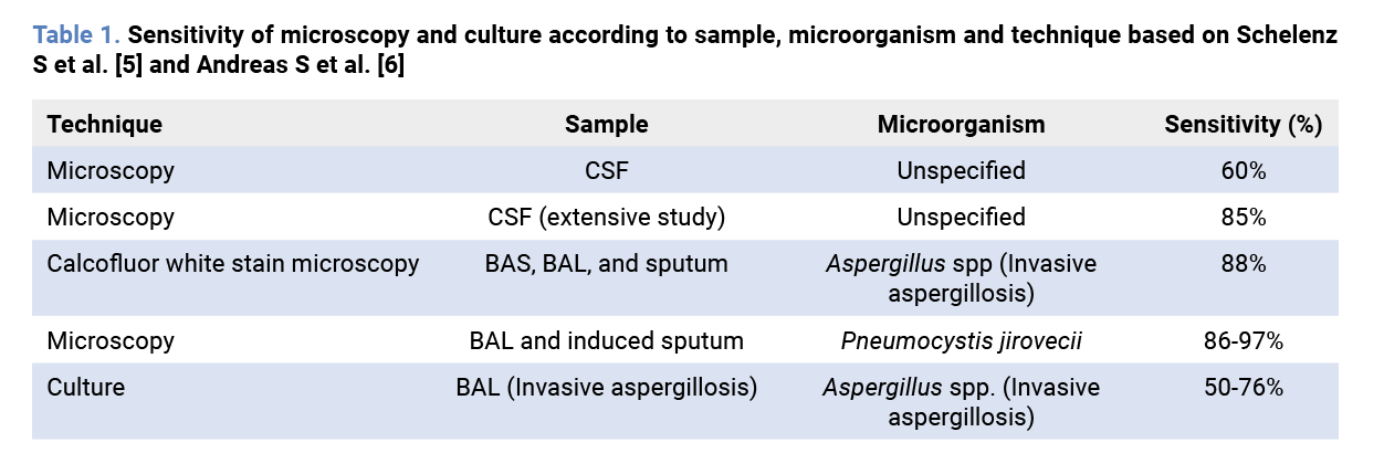 In relation to invasive aspergillosis (IA), the isolation of Aspergillus spp. from lower respiratory tract is the first indication of IA. However, the probability of a non-selected risk patient from whom Aspergillus spp. has been isolated having IA was calculated to be 12%. However, this diagnostic yield is influenced by underlying comorbidities: 72% in patients with neutropenia, 58% in solid organ transplant recipients, and 28% in critically ill patients without those risk factors [4].
Currently, several non-classical diagnostic techniques are available for the detection of invasive fungal infections (IFIs), primarily based on the identification of specific fungal biomarkers. These include: 1,3-®-D-glucan panfungal antigen (BDG), Aspergillus spp. antigen (galactomannan, GAL), lateral flow for antigen/antibodies detection, immunochromatography, Candida albicans, germ tube antibodies (CAGTA), manano antigen/antibodies (Mn/Ab Mn) and nuclear acid amplification tests (NAAT) The selection of a specific test depends largely on the suspected fungal pathogen, such as Candida spp., Aspergillus spp., Pneumocystis jirovecii, or organisms from the order Mucorales. In all cases, the pre-test probability is a critical factor in guiding both the choice of diagnostic method and the interpretation of results. According to Fagan’s application of Bayes’ theorem to diagnostic reasoning, the positive likelihood ratio indicates how much a positive test result increases the probability that the patient truly has the disease, by comparing the probability of a true positive to that of a false positive [7].
Invasive Candidiasis (IC). Blood cultures remain the gold standard for the diagnosis of candidemia. According to the 2025 guidelines [8], biomarkers and nucleic acid amplification tests (NAATs) are not yet routinely recommended as standalone tools for diagnosing invasive candidiasis (IC). However, they provide valuable support in both diagnosis and prognosis.
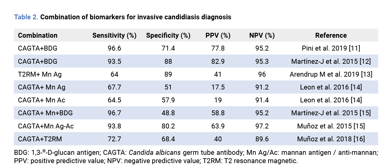
Available biomarkers include (1→3)-β-D-glucan (BDG), a panfungal marker; the detection of mannan antigen and anti-mannan antibodies (Mn/Ab Mn); and the Candida albicans germ tube antibody (CAGTA), which targets cell wall components of Candida spp. The sensitivity and specificity of these techniques vary between 60–87% and 58–90%, respectively (Table 2).
 Several commercial NAAT platforms are available for Candida spp. detection, with reported sensitivities ranging from 60% to 95% and specificities between 92% and 99%. Among them, the T2Candida® magnetic resonance platform (T2 Biosystems®, Lexington, Massachusetts, USA) was notable as the only FDA-approved molecular assay for candidemia, with sensitivity and specificity rates around 90–95%. Unfortunately, this technology has recently been withdrawn from the market.
The main advantage of combining biomarkers and molecular tests lies in their ability to increase the negative predictive value (NPV) for IC. Conversely, their positive predictive value (PPV) improves—either individually or in combination—as the pretest probability of IC increases (Table 2).
Among all biomarkers, BDG is the most widely used in clinical practice. Nevertheless, evidence regarding its utility for guiding treatment decisions or antifungal discontinuation in critically ill patients remains conflicting [9,10]. Further multicentre studies are warranted to clarify its role and inform clinical management in this population.
Invasive Aspergillosis (IA). IA primarily occurs in patients with significant comorbidities, including moderate to severe chronic obstructive pulmonary disease (COPD), decompensated liver cirrhosis, uncontrolled HIV infection, and solid tumors. Additional risk factors include prolonged or high-dose corticosteroid therapy, invasive procedures in the ICU, prolonged antibiotic treatment, and recent or concurrent viral infections [17].
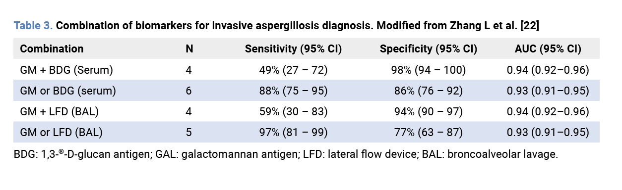
Several diagnostic tools are available for the detection of IA, including biomarkers such as GAL (Aspergillus-specific) and BDG (nonspecific), as well as rapid assays like the GAL or JF5 lateral flow assays, with high accordance with ELISA test, and NAATs [18,19]. A metanalysis showed a serum GAL sensitivity (11% IA prevalence, Platelia Bio-Rad® cut-off: 1) of 72% in neutropenic population [20]. However, this sensitivity decreases to 16.7% in non-neutropenic population [21]. In contrast, its use in bronchoalveolar lavage (BAL) demonstrates higher sensitivity, around 60–90% [18,21]. Notably, the combined use of GAL in BAL and serum BDG significantly enhances diagnostic performance, with sensitivity and NPV reaching up to 100% for IA in critical ill population [18]. However, the addition of NAAT tests to this combination does not result in further improvement in PPV or specificity [18] (Table 3).
 Pneumocystis jirovecii pneumonia (PJP). PJP primarily occurs in patients with underlying risk factors such as immunosuppression, prolonged corticosteroid therapy, respiratory comorbidities (i.e COPD), sepsis, use of novel molecular inhibitors, advanced age, and underlying immunodeficiencies such as HIV infection. Although its true incidence remains unknown, PJP is increasingly recognized as an emerging infection, with reported hospital outbreaks and a growing number of cases in non-traditional risk groups [17].
The gold standard for diagnosing PJP remains immunofluorescence microscopy on respiratory tract specimens. However, NAAT (primarily quantitative RT–PCR) is playing an increasingly prominent role in the diagnostic approach. When performed on BAL samples, NAAT offers near 100% sensitivity and a high NPV, making it a valuable tool to rule out PJP infection. The use of quantitative cut-off values to distinguish between colonization and true infection has been proposed. However, no consensus has been reached to date, primarily due to the lack of standardization across diagnostic techniques [23].
Serum BDG levels are typically elevated in PJP, but BDG alone is insufficient to definitively rule out the disease [24]. Non-invasive specimens such as nasopharyngeal aspirates and oral rinses are now commonly used for NAAT, particularly in patients with a high pretest probability. In such cases, the combined use of BDG and NAAT can support the PJP diagnosis. However, a negative result from both tests does not definitively exclude PJP infection [23].
Mucormycosis. The diagnosis of mucormycosis remains a significant challenge due to the absence of specific biomarkers and reliable non-invasive tests [17]. Currently, the detection of mucorales by NAAT in serum for the non-invasive diagnosis of mucormycosis is gaining importance, as reflected in the MODIMUCOR trial [25]. Multicenter reproducibility studies are being conducted in the following areas. Recently, an immunochromatographic assay have been developed using a monoclonal antibody targeting Rhizopus arrizhus (the most common causative species of mucormycosis in Europe) or panmucoral for the detection of a hyphal growth–associated polysaccharide in both BAL and serum samples [26,27].
Conclusion
The diagnosis of invasive fungal infections requires a multifaceted approach that begins with a thorough clinical history and an accurate assessment of pretest probability. Routine use of direct microscopy and culture remains essential as a first diagnostic step. Understanding local prevalence, epidemiological patterns, and individual risk factors is key to guiding both clinical suspicion and test selection. Given the variability in diagnostic performance across fungal pathogens, it is crucial to choose the most appropriate test based on the suspected organism. In general, available diagnostic tools offer higher NPVs, making them especially valuable for ruling out infection. When possible, combining different diagnostic tests can significantly improve sensitivity and overall diagnostic accuracy, ultimately leading to better patient outcomes. Funding None to declare. Conflict of interest The authors declare no conflict of interest. Author contributions A.M.B.; writing—original draft preparation, I.Q.M writing-original draft preparation, J.G.R; writing—review and editing. All authors have read and agreed to the published version of the manuscript.References
- Denning DW. Global incidence and mortality of severe fungal disease. Lancet Infect Dis 2024;24:e428–38. Available from: https://doi.org/10.1016/S1473-3099(23)00692-8
- Lass-Flörl C, Steixner S. The changing epidemiology of fungal infections. Mol Aspects Med. 2023;94:101215. Available from: https://doi.org/10.1016/j.mam.2023.101215
- Spanish Society of Critical Intensive Care Medicine and Coronary Units, Vall d’Hebron University Hospital. National Surveillance Study of Nosocomial Infection in ICU - Hospitals in Europe Link for Infection Control through Surveillance (ENVIN-HELICS) n.d. Available from: https://hws.vhebron.net/ENVIN-helics/
- Guinea J, Torres-Narbona M, Gijón P, Muñoz P, Pozo F, Peláez T, et al. Pulmonary aspergillosis in patients with chronic obstructive pulmonary disease: incidence, risk factors, and outcome. Clin Microbiol Infect 2010;16:870–7. Available from: https://doi.org/10.1111/j.1469-0691.2009.03015.x
- Schelenz S, Barnes RA, Barton RC, Cleverley JR, Lucas SB, Kibbler CC, et al. British Society for Medical Mycology best practice recommendations for the diagnosis of serious fungal diseases. Lancet Infect Dis 2015;15:461–74. Available from: https://doi.org/10.1016/S1473-3099(15)70006-X
- Andreas S, Heindl S, Wattky C, Möller K, Rüchel R. Diagnosis of pulmonary aspergillosis using optical brighteners. Eur Respir J 2000;15:407. Available from: https://doi.org/10.1034/j.1399-3003.2000.15b30.x
- Fagan TJ. Nomogram for Bayes’s Theorem. New Engl J Med 1975;293:257. Available from: https://doi.org/10.1056/NEJM197507312930513
- Cornely OA, Sprute R, Bassetti M, Chen SC-A, Groll AH, Kurzai O, et al. Global guideline for the diagnosis and management of candidiasis: an initiative of the ECMM in cooperation with ISHAM and ASM. Lancet Infect Dis 2025;25:e280–93. Available from: https://doi.org/10.1016/S1473-3099(24)00749-7
- Nucci M, Nouér SA, Esteves P, Guimarães T, Breda G, De Miranda BG, et al. Discontinuation of empirical antifungal therapy in ICU patients using 1,3-β-d-glucan. J Antimicrob Chemother 2016;71:2628–33. Available from: https://doi.org/10.1093/jac/dkw188
- Christner M, Abdennadher B, Wichmann D, Kluge S, Pepić A, Aepfelbacher M, et al. The added value of (1,3)-β-D-glucan for the diagnosis of Invasive Candidiasis in ICU patients: a prospective cohort study. Infection 2024;52:73–81. Available from: https://doi.org/10.1007/s15010-023-02053-4
- Pini P, Venturelli C, Girardis M, Forghieri F, Blasi E. Prognostic Potential of the Panfungal Marker (1 → 3)-β-d-Glucan in Invasive Mycoses Patients. Mycopathologia 2019;184:147–50. Available from: https://doi.org/10.1007/s11046-018-0282-5
- Martínez-Jiménez MC, Muñoz P, Valerio M, Alonso R, Martos C, Guinea J, et al. Candida biomarkers in patients with candidaemia and bacteraemia. J Antimicrob Chemother 2015;70:2354–61. Available from: https://doi.org/10.1093/jac/dkv090
- Arendrup MC, Andersen JS, Holten MK, Krarup KB, Reiter N, Schierbeck J, et al. Diagnostic Performance of T2Candida Among ICU Patients With Risk Factors for Invasive Candidiasis. Open Forum Infect Dis 2019;6:ofz136. Available from: https://doi.org/10.1093/ofid/ofz136
- The Cava Trem Study Group, León C, Ruiz-Santana S, Saavedra P, Castro C, Loza A, et al. Contribution of Candida biomarkers and DNA detection for the diagnosis of invasive candidiasis in ICU patients with severe abdominal conditions. Crit Care 2016;20:149. Available from: https://doi.org/10.1186/s13054-016-1324-3
- Martínez-Jiménez MC, Muñoz P, Guinea J, Valerio M, Alonso R, Escribano P, et al. Potential role of Candida albicans germ tube antibody in the diagnosis of deep-seated candidemia. Med Mycol 2014;52:270–5. Available from: https://doi.org/10.1093/mmy/myt025
- Muñoz P, Vena A, Machado M, Gioia F, Martínez-Jiménez MC, Gómez E, et al. T2Candida MR as a predictor of outcome in patients with suspected invasive candidiasis starting empirical antifungal treatment: a prospective pilot study. J Antimicrob Chemother 2018;73:iv6–12. Available from: https://doi.org/10.1093/jac/dky047
- Donnelly JP, Chen SC, Kauffman CA, Steinbach WJ, Baddley JW, Verweij PE, et al. Revision and Update of the Consensus Definitions of Invasive Fungal Disease From the European Organization for Research and Treatment of Cancer and the Mycoses Study Group Education and Research Consortium. Clin Infect Dis. 2020;71:1367–76. Available from: https://doi.org/10.1093/cid/ciz1008
- Boch T, Reinwald M, Spiess B, Liebregts T, Schellongowski P, Meybohm P, et al. Detection of invasive pulmonary aspergillosis in critically ill patients by combined use of conventional culture, galactomannan, 1-3-beta-D-glucan and Aspergillus specific nested polymerase chain reaction in a prospective pilot study. J Crit Care 2018;47:198–203. Available from: https://doi.org/10.1016/j.jcrc.2018.07.001
- Wan L, Cai X, Ling M, Kan J, Yin M, Wang H. Evaluation of the JF5-based Aspergillus galactomannoprotein lateral flow device for diagnosing invasive aspergillosis in cancer patients. Eur J Clin Microbiol Infect Dis 2024;43:1221–9. Available from: https://doi.org/10.1007/s10096-024-04830-x
- Leeflang MM, Debets-Ossenkopp YJ, Wang J, Visser CE, Scholten RJ, Hooft L, et al. Galactomannan detection for invasive aspergillosis in immunocompromised patients. Cochrane Database Syst Rev. 2015;2017. Available from: https://doi.org/10.1002/14651858.CD007394.pub2
- Wu Z, Wang L, Tan L, Wu J, Chen Z, Hu M. Diagnostic value of galactomannan in serum and bronchoalveolar lavage fluid for invasive pulmonary aspergillosis in non-neutropenic patients. Diagn Microbiol Infect Dis 2021;99:115274. Available from: https://doi.org/10.1016/j.diagmicrobio.2020.115274
- Zhang L, Guo Z, Xie S, Zhou J, Chen G, Feng J, et al. The performance of galactomannan in combination with 1,3-β-D-glucan or aspergillus-lateral flow device for the diagnosis of invasive aspergillosis: Evidences from 13 studies. Diagn Microbiol Infect Dis. 2019;93:44–53. Available from: https://doi.org/10.1016/j.diagmicrobio.2018.08.005
- Guegan H, Robert-Gangneux F. Molecular diagnosis of Pneumocystis pneumonia in immunocompromised patients. Curr Opin Infect Dis. 2019;32:314–21. Available from: https://doi.org/10.1097/QCO.0000000000000559
- Del Corpo O, Butler-Laporte G, Sheppard DC, Cheng MP, McDonald EG, Lee TC. Diagnostic accuracy of serum (1-3)-β-D-glucan for Pneumocystis jirovecii pneumonia: a systematic review and meta-analysis. Clin Microbiol Infect 2020;26:1137–43. Available from: https://doi.org/10.1016/j.cmi.2020.05.024
- Millon L, Caillot D, Berceanu A, Bretagne S, Lanternier F, Morio F, et al. Evaluation of Serum Mucorales Polymerase Chain Reaction (PCR) for the Diagnosis of Mucormycoses: The MODIMUCOR Prospective Trial. Clin Infect Dis 2022;75:777–85. Available from: https://doi.org/10.1093/cid/ciab1066
- Rousselot J, Millon L, Scherer E, Bourgeois N, Imbert S, Dupont D, et al. Detection of Mucorales antigen in bronchoalveolar lavage samples using a newly developed lateral-flow device. J Clin Microbiol 2025:e00226-25. Available from: https://doi.org/10.1128/jcm.00226-25
- Davies GE, Thornton CR. Development of a Monoclonal Antibody and a Serodiagnostic Lateral-Flow Device Specific to Rhizopus arrhizus (Syn. R. oryzae), the Principal Global Agent of Mucormycosis in Humans. JoF 2022;8:756. Available from: https://doi.org/10.3390/jof8070756
Explore
Articles
Volumes
About the journal
Most read
Recomendaciones para la prevención y el tratamiento de las infecciones respiratorias en adultos mayores institucionalizados
Revista Española de Quimioterapia
Acute infectious gastroenteritis in childhood: the role of rapid multiplex molecular syndromic panels in diagnosis and clinical management
Revista Española de Quimioterapia
Características epidemiológicas de pacientes con aislamientos de Enterobacterales productores de carbapenemasas-NDM en un hospital de alta complejidad
Revista Española de Quimioterapia
Review on the management of intra-abdominal candidiasis
Revista Española de Quimioterapia
Neoscytalidium dimidiatum: a rare locally acquired deep cutaneous infection in a renal transplant recipient
Revista Española de Quimioterapia