Case report
June 2025, Volume 38, nº 3
7 min
Disseminated histoplasmosis with bone marrow infiltration in a newly diagnosed HIV patient
Emilio Guirao-Arrabal1,* , Natalia Chueca-Porcuna2 , Elena Cornejo-Calvo3 , Laura Alcázar-Fuoli4 , Andrés Ruiz-Sancho1
1Infectious Diseases Unit, Hospital San Cecilio, Granada, Spain
2Microbiology Unit, Hospital San Cecilio, Granada, Spain
3Hematology Unit, Hospital San Cecilio, Granada, Spain
4Mycology Reference Lab, National Centre for Microbiology, Instituto de Salud Carlos III, Majadahonda, Madrid, Spain
http://www.doi.org/10.37201/req/008.2025
Receipt Date:
09/01/2025
Acceptance Date:
02/04/2025
Publication Date:
22/04/2025
How to cite
Guirao-Arrabal E, Chueca-Porcuna N, Cornejo-Calvo E, Alcázar-Fuoli L, Ruiz-Sancho A. Disseminated histoplasmosis with bone marrow infiltration in a newly diagnosed HIV patient. Rev Esp Quimioter. 2025; 38(3): 249-52. Available from: http://www.doi.org/10.37201/req/008.2025
Metrics
542
Views
44
Downloads
Share
Share
Sir,
A 41-year-old male patient experienced unintended weight loss of 8 kg for several months and progressive vomiting. The patient was born in Colombia but had been living in Spain for three years (did not travel to Colombia during this 3-year period), and he was not aware of any pre-existing conditions. He had no occupational or recreational history of environmental exposure to organic or inorganic hazardous agents. His blood pressure was 115/68 mmHg, heart rate 89 bpm, 96% oxygen saturation on room air, and he was afebrile. Cardiorespiratory auscultation was unremarkable. Abdominal examination only revealed mild hepatomegaly, and no peripheral lymphadenopathy was detected. An initial blood test in the emergency department revealed Hb 11.8 g/dL, 82,000 platelets/mcL, elevated ferritin (2,886 ng/mL), high LDH (731 U/L), low haptoglobin (17 mg/dL), C-reactive protein of 83 mg/L and procalcitonin at 0.19 ng/ml. Human immunodeficiency virus (HIV) serology was positive and he was admitted to our Infectious Diseases Unit for further study and treatment.
Aerobic, anaerobic and mycobacterial blood cultures were negative. HIV viral load of 216,000 copies/mL and CD4-T lymphocytes count was 67/mcL (13%). Both beta-D-glucan and galactomannan in a blood sample were positive (with values of 8.8 pg/mL and 5.9 mcg/L respectively). Serum Cryptococcal antigen, Quantiferon-TB Gold Plus® (interferon-gamma release assay or IGRA test), and hepatitis B, hepatitis C and Treponema pallidum serologies were all negative. Toxoplasma IgG, CMV IgG and hepatitis A IgG serologies were all positive. CMV serum viral load was 38 copies/mL. EBV viral load was not performed.
A chest X-ray was normal. A chest CT showed a 15 mm short-axis right hilar lymphadenopathy, a 6 mm right apical calcified nodule, and a 3 mm nodule in the middle lobe. Due to the poor quality of the sputum, neither culture nor molecular techniques could be performed in any respiratory sample. A bone marrow aspirate was performed. The smear is shown in Figure 1.
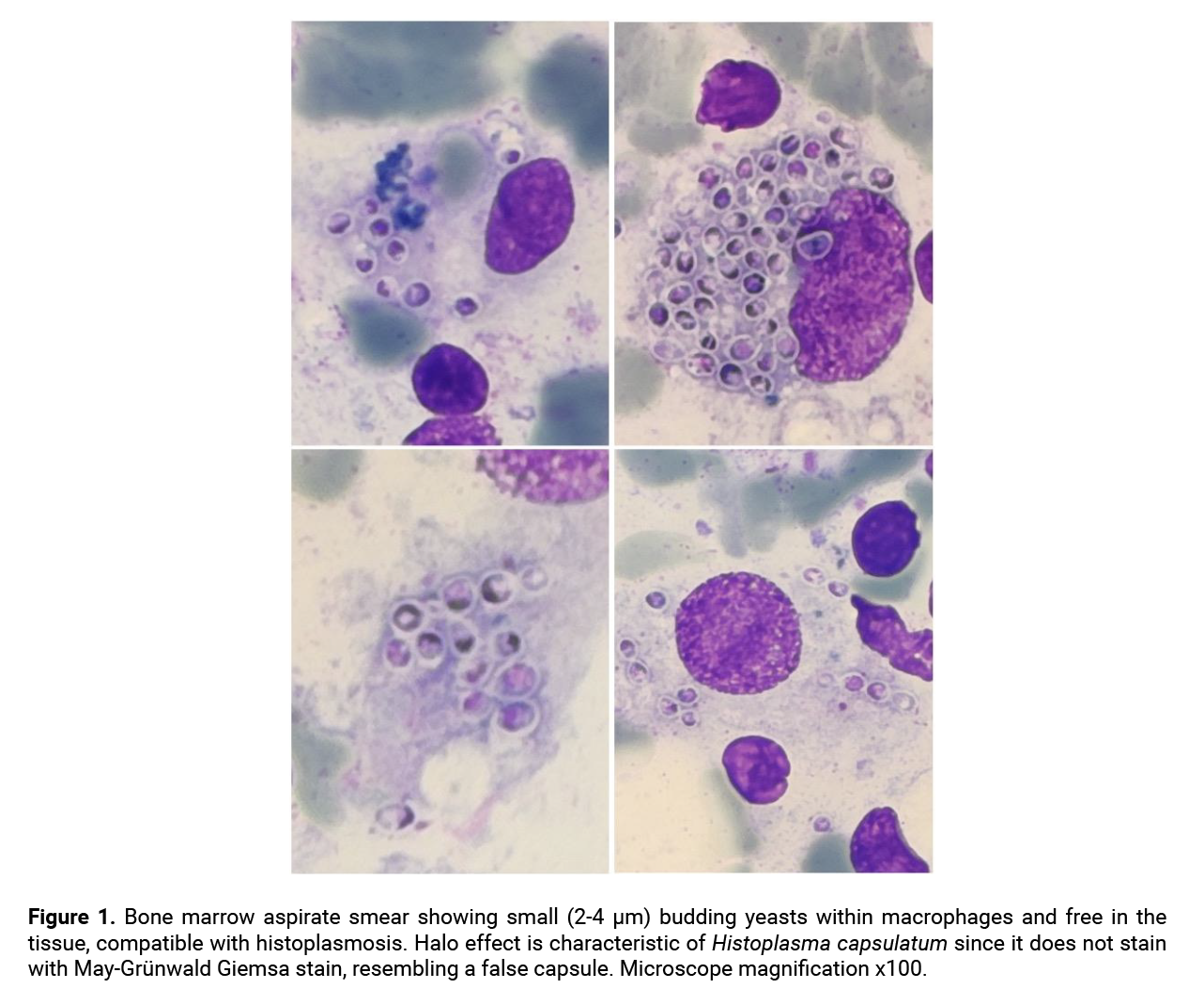 Based on clinical suspicion and the visualization of structures highly compatible with histoplasmosis, the patient was started on treatment, pending definitive results: liposomal amphotericin B (L-AmB) 4 mg/kg/24h to treat a possible histoplasmosis, as well as bictegravir/emtricitabine/tenofovir alafenamide (for HIV infection) and cotrimoxazole prophylaxis (to prevent Pneumocystis jirovecii pneumonia) were initiated. The patient’s general condition improved, and cytopenias began to resolve rapidly under antifungal therapy.
Leishmania spp and Parvovirus B19 PCR were performed in the bone marrow sample, and both were negative.
Bone marrow samples were sent to the National Center for Microbiology in Madrid, for further microbiological analysis. Culture on Sabouraud dextrose agar at 25-30°C from bone marrow aspirate sample revealed fungal structures, including tuberculate macroconidia with thick walls, surface projections and smooth-surfaced microconidia, compatible with Histoplasma capsulatum. In this culture sample, DNA extraction and molecular identification of H. capsulatum were performed by amplifying and sequencing the rDNA internal transcribed spacer region and the result was positive. Neither blood PCR for H. capsulatum nor urinary antigen tests were performed.
H. capsulatum var. capsulatum is a dimorphic fungus found in many parts of the world, mainly in humid climate regions (including countries in America, Africa, and Asia as well as some parts of central Europe, though not in Spain) [1]. Approximately 32% of histoplasmin skin test positivity has been reported throughout Latin America, with important differences between regions [2]. This implies that a significant proportion of the population from endemic countries has been previously infected and is susceptible to reactivation through any type of immunosuppression (HIV, solid organ transplantation, anti-TNF, etc.).
Primary infection is acquired via inhalation of microconidia from soil contaminated with bird or bat guano. Histoplasmosis can develop as an acute infection (usually asymptomatic, self-limited fever or pneumonia in both immunocompromised and immunocompetent individuals), reactivation (in which immunocompromised patients may develop disseminated disease via this etiopathogenic route) or reinfection. Immunity to H. capsulatum is mediated by INF-gamma, produced by macrophages and CD4 T lymphocytes, so severely immunocompromised patients, like those with advanced HIV, are prone to severe disease. Disseminated disease forms develop over 2-4 weeks and can produce a wide variety of symptoms: fever, fatigue, weight loss, shortness of breath, diarrhea with or without bleeding, and skin eruptions. At physical examination, some signs are common: lymphadenopathies, hepatomegaly or splenomegaly, skin or oral lesions ranging from maculopapular eruptions to ulcerations.
Pancytopenia, transaminitis and high LDH are common laboratory findings [3]. Hemophagocytic lymphohistiocytosis, disseminated intravascular coagulation and septic shock have been associated with histoplasmosis [3–5].
Serological testing lacks sensitivity in HIV patients. Diagnosis relies on culture and molecular techniques; however, culture takes 2 to 8 weeks to yield results [6]. Samples for culture or PCR can be obtained from bone marrow, blood, sputum/bronchoscopy, or CSF, each with different sensitivities. Where available, histoplasma antigen testing can accelerate diagnosis. This latter test can be performed on urine, blood, bronchoalveolar lavage fluid, or cerebrospinal fluid. Serum biomarkers like beta-glucan and galactomannan can both be positive, but there is limited data regarding the sensitivity and specificity of these serological markers [7]. It would be interesting to know their role as a first step when bone marrow aspiration cannot be performed early, and the patient is unstable. When antifungal treatment must be promptly initiated, a serum sample can be obtained first for fungal biomarkers (beta-D-glucan and galactomannan), so a positive result can later support the diagnosis of histoplasmosis.
We followed the classical liposomal amphotericin-B regimen, which is dosed as 3-5 mg/kg/day for 1-2 weeks, followed by oral itraconazole 200 mg twice daily for at least 12 months [8]. A single 10 mg/kg L-AmB induction dose (followed by oral itraconazole therapy) may be sufficient to achieve clinical and microbiological cure, based on a recent phase II clinical trial [9].
This case report emphasizes the role of early bone marrow aspiration in HIV patients with cytopenias and low CD4 counts. This diagnostic technique can lead to a final diagnosis in other infections like visceral leishmaniasis, disseminated MAC infection or CMV. Histoplasmosis is an uncommon opportunistic infection in Spain but should be taken into consideration in those patients from endemic regions. The increase in international travels may cause asymptomatic infections in individuals from non-endemic countries, which could later reactivate under immunosuppressive conditions. We aim to highlight this opportunistic fungal infection as a potentially life-threatening condition that is rarely diagnosed in Spain. However, it should be considered, given the evolving epidemiology of infectious diseases driven by migration, climate change, and tourism.
Funding
None to declare.
Conflict of interest
Authors declare no conflicts of interest.
Author contributions
Conceptualization, E. G-A. and N. C-P.; methodology, E. G-A.; resources, N. C-P.; writing—original draft preparation, E. G-A., E. C-C. and L. A-F.; writing—review and editing, E. G-A. and A. R-S.; image acquisition: E. C-C.; supervision, E. G-A. All authors have read and agreed to the published version of the manuscript.
Based on clinical suspicion and the visualization of structures highly compatible with histoplasmosis, the patient was started on treatment, pending definitive results: liposomal amphotericin B (L-AmB) 4 mg/kg/24h to treat a possible histoplasmosis, as well as bictegravir/emtricitabine/tenofovir alafenamide (for HIV infection) and cotrimoxazole prophylaxis (to prevent Pneumocystis jirovecii pneumonia) were initiated. The patient’s general condition improved, and cytopenias began to resolve rapidly under antifungal therapy.
Leishmania spp and Parvovirus B19 PCR were performed in the bone marrow sample, and both were negative.
Bone marrow samples were sent to the National Center for Microbiology in Madrid, for further microbiological analysis. Culture on Sabouraud dextrose agar at 25-30°C from bone marrow aspirate sample revealed fungal structures, including tuberculate macroconidia with thick walls, surface projections and smooth-surfaced microconidia, compatible with Histoplasma capsulatum. In this culture sample, DNA extraction and molecular identification of H. capsulatum were performed by amplifying and sequencing the rDNA internal transcribed spacer region and the result was positive. Neither blood PCR for H. capsulatum nor urinary antigen tests were performed.
H. capsulatum var. capsulatum is a dimorphic fungus found in many parts of the world, mainly in humid climate regions (including countries in America, Africa, and Asia as well as some parts of central Europe, though not in Spain) [1]. Approximately 32% of histoplasmin skin test positivity has been reported throughout Latin America, with important differences between regions [2]. This implies that a significant proportion of the population from endemic countries has been previously infected and is susceptible to reactivation through any type of immunosuppression (HIV, solid organ transplantation, anti-TNF, etc.).
Primary infection is acquired via inhalation of microconidia from soil contaminated with bird or bat guano. Histoplasmosis can develop as an acute infection (usually asymptomatic, self-limited fever or pneumonia in both immunocompromised and immunocompetent individuals), reactivation (in which immunocompromised patients may develop disseminated disease via this etiopathogenic route) or reinfection. Immunity to H. capsulatum is mediated by INF-gamma, produced by macrophages and CD4 T lymphocytes, so severely immunocompromised patients, like those with advanced HIV, are prone to severe disease. Disseminated disease forms develop over 2-4 weeks and can produce a wide variety of symptoms: fever, fatigue, weight loss, shortness of breath, diarrhea with or without bleeding, and skin eruptions. At physical examination, some signs are common: lymphadenopathies, hepatomegaly or splenomegaly, skin or oral lesions ranging from maculopapular eruptions to ulcerations.
Pancytopenia, transaminitis and high LDH are common laboratory findings [3]. Hemophagocytic lymphohistiocytosis, disseminated intravascular coagulation and septic shock have been associated with histoplasmosis [3–5].
Serological testing lacks sensitivity in HIV patients. Diagnosis relies on culture and molecular techniques; however, culture takes 2 to 8 weeks to yield results [6]. Samples for culture or PCR can be obtained from bone marrow, blood, sputum/bronchoscopy, or CSF, each with different sensitivities. Where available, histoplasma antigen testing can accelerate diagnosis. This latter test can be performed on urine, blood, bronchoalveolar lavage fluid, or cerebrospinal fluid. Serum biomarkers like beta-glucan and galactomannan can both be positive, but there is limited data regarding the sensitivity and specificity of these serological markers [7]. It would be interesting to know their role as a first step when bone marrow aspiration cannot be performed early, and the patient is unstable. When antifungal treatment must be promptly initiated, a serum sample can be obtained first for fungal biomarkers (beta-D-glucan and galactomannan), so a positive result can later support the diagnosis of histoplasmosis.
We followed the classical liposomal amphotericin-B regimen, which is dosed as 3-5 mg/kg/day for 1-2 weeks, followed by oral itraconazole 200 mg twice daily for at least 12 months [8]. A single 10 mg/kg L-AmB induction dose (followed by oral itraconazole therapy) may be sufficient to achieve clinical and microbiological cure, based on a recent phase II clinical trial [9].
This case report emphasizes the role of early bone marrow aspiration in HIV patients with cytopenias and low CD4 counts. This diagnostic technique can lead to a final diagnosis in other infections like visceral leishmaniasis, disseminated MAC infection or CMV. Histoplasmosis is an uncommon opportunistic infection in Spain but should be taken into consideration in those patients from endemic regions. The increase in international travels may cause asymptomatic infections in individuals from non-endemic countries, which could later reactivate under immunosuppressive conditions. We aim to highlight this opportunistic fungal infection as a potentially life-threatening condition that is rarely diagnosed in Spain. However, it should be considered, given the evolving epidemiology of infectious diseases driven by migration, climate change, and tourism.
Funding
None to declare.
Conflict of interest
Authors declare no conflicts of interest.
Author contributions
Conceptualization, E. G-A. and N. C-P.; methodology, E. G-A.; resources, N. C-P.; writing—original draft preparation, E. G-A., E. C-C. and L. A-F.; writing—review and editing, E. G-A. and A. R-S.; image acquisition: E. C-C.; supervision, E. G-A. All authors have read and agreed to the published version of the manuscript.
References
- Kontogiannis D, Di Lorenzo A, Zaçe D, Benvenuto D, Moccione M, Muratore G, et al. Histoplasmosis in patients living with HIV in Europe: review of literature. Front Microbiol 2024;15:1–12. https://doi.org/10.3389/fmicb.2024.1418530.
- Adenis AA, Valdes A, Cropet C, McCotter OZ, Derado G, Couppie P, et al. Burden of HIV-associated histoplasmosis compared with tuberculosis in Latin America: a modelling study. Lancet Infect Dis 2018;18:1150–9. https://doi.org/10.1016/S1473-3099(18)30354-2.
- Manuel C, Adamian C, Lima MA De, Adler A, Martins F, Cardoso M, et al. Progressive disseminated histoplasmosis in HIV-positive patients 2022. https://doi.org/10.1177/09564624221076605.
- Cooper JL, Spring AM, Low DJ. Disseminated intravascular coagulation from disseminated histoplasmosis in uncontrolled HIV – Case report. IDCases 2021;26:e01270. https://doi.org/10.1016/j.idcr.2021.e01270.
- Huang AM. Hemophagocytic lymphohistiocytosis and disseminated histoplasmosis. Blood 2014;123:2449–2449. https://doi.org/10.1182/blood-2014-01-550657.
- Araúz AB, Papineni P. Histoplasmosis. Infect Dis Clin North Am 2021;35:471–91. https://doi.org/10.1016/j.idc.2021.03.011.
- Giacobbe DR, Bono V Del, Viscoli C, Giacobbe DR, Bono V Del, Viscoli C, et al. Expert Review of Anti-infective Therapy Use of 1, 3- β -D-glucan in invasive fungal diseases in hematology patients Use of 1, 3- β -D-glucan in invasive fungal diseases in hematology patients. Expert Rev Anti Infect Ther 2017;15:1101–12. https://doi.org/10.1080/14787210.2017.1401467.
- Eichenberger EM, Little JS, Baddley JW. Histoplasmosis. Infect Dis Clin North Am 2024. https://doi.org/10.1016/j.idc.2024.11.009.
- Pasqualotto AC, Lana DD, Godoy CSM, Leitão TDMJS, Bay MB, Damasceno LS, et al. Single High Dose of Liposomal Amphotericin B in Human Immunodeficiency Virus/AIDS-Related Disseminated Histoplasmosis: A Randomized Trial. Clinical Infectious Diseases 2023;77:1126–32. https://doi.org/10.1093/cid/ciad313.
Explore
Articles
Volumes
About the journal
Most read
Recomendaciones para la prevención y el tratamiento de las infecciones respiratorias en adultos mayores institucionalizados
Revista Española de Quimioterapia
Acute infectious gastroenteritis in childhood: the role of rapid multiplex molecular syndromic panels in diagnosis and clinical management
Revista Española de Quimioterapia
Características epidemiológicas de pacientes con aislamientos de Enterobacterales productores de carbapenemasas-NDM en un hospital de alta complejidad
Revista Española de Quimioterapia
Review on the management of intra-abdominal candidiasis
Revista Española de Quimioterapia
Neoscytalidium dimidiatum: a rare locally acquired deep cutaneous infection in a renal transplant recipient
Revista Española de Quimioterapia