Letter
June 2026, Volume 39, nº 3
9 min
Multiple intracranial infectious aneurysms in a case of endocarditis. Intravascular management
Múltiples aneurismas infecciosos intracraneales en un caso de endocarditis. Manejo endovascular
Monique Boukobza1*  , Sylvie Lariven2 , Jean-Pierre Laissy1,3,4
1Department of Radiology, Bichat-Claude Bernard Hospital, Assistance Publique-Hôpitaux de Paris, Paris, France
2Infectious and Tropical Diseases Department, Bichat-Claude Bernard Hospital, Assistance Publique-Hôpitaux de Paris, Paris, France
3INSERM U1148, Paris, France
4Université Paris Cité, Paris, France
*Correspondence:
Monique Boukobza. m.boukobza@orange.fr
Receipt Date:
24/12/2025
Acceptance Date:
27/03/2026
Publication Date:
30/04/2026
How to cite
Boukobza M, Lariven S, Laissy J-P. Multiple intracranial infectious aneurysms in a case of endocarditis. Intravascular management. Rev Esp Quimioter 2026; 39(3):266-269. Available from: http://www.doi.org/10.37201/req/139.2025
Metrics
658
Views
21
Downloads
Share
Share
Sir,
Infective endocarditis (IE) due to Abiotrophia spp. and Granulicatella spp., previously described as nutritionally variant Streptococcus spp., represent between 1–3% of all IE cases. Intracranial infectious aneurysms (IIAs) associated with Abiotrophia defectiva remain poorly understood due to the limited number of reported cases caused by this species [1-9]. We report a patient with six unruptured IIAs in IE caused by A. defectiva.
A 52-year-old obese man (body mass index: 32.5), with a history of arterial hypertension, mitral valve (MV) posterior leaflet (P2) prolapse with mitral regurgitation, and hepatic steatosis (50%), presented with a 2-month history of fatigue, anorexia, and weight loss of 4 kg. Examination was unremarkable. Considering his history and elevated inflammatory markers, IE was suspected. The initial report on blood cultures suspected streptococci at Gram stain.
Treatment with amoxicillin (12 g/day) plus ceftriaxone (2 g/day) was initiated.
A transthoracic/transesophageal echocardiogram (TTE/TEE) showed a 4mm vegetation on the MV and severe MV insufficiency due to P2 prolapsus. The first blood culture revealed Abiotrophia defectiva. The antibiotic treatment was switched to ceftriaxone (2 g/day) plus gentamicin (3 mg/kg/day) for two weeks and then ceftriaxone alone for 4 weeks. Speciation of Abiotrophia defectiva was performed using 16S rRNA sequencing of the blood culture isolate. The antibiotics were stopped at six weeks, and conservative valve treatment was the single option.
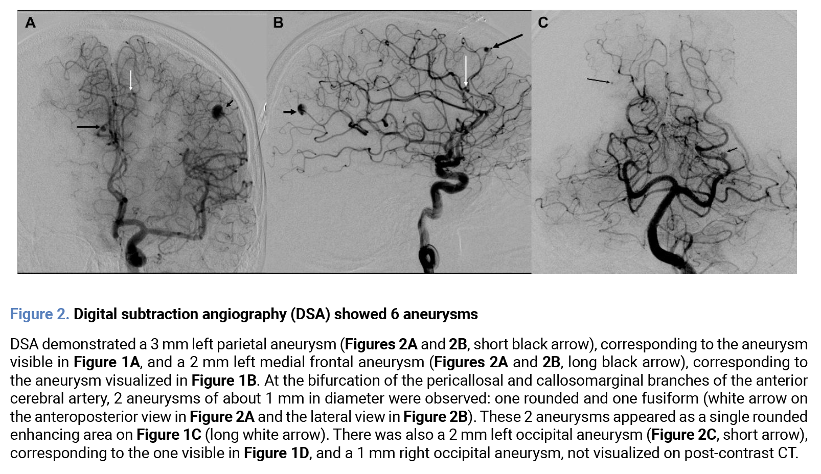
Whole-body and cranial CT angiography (CTA), as part of the initial IE workup at our institution, performed at hospital day (HD) 2, revealed 4 IIAs without intracranial hemorrhage or ischemic lesions (Figure 1A-D) and multiple splenic infarcts. High-definition digital subtraction angiography (DSA) at HD 4 showed a total of six IIAs. Three were treated by exclusion of the parent artery by glue embolization (n=1, 3 mm, parietal branch of the left middle cerebral artery (MCA) (Figures 2A and B), by coils (n=1, 1.5mm, anterior cerebral artery (ACA) frontal branch), and by both glue and coil embolization (n=1, 2 mm, from a branch of the left occipital artery, arising from the left MCA; Figure 2C, short arrow).
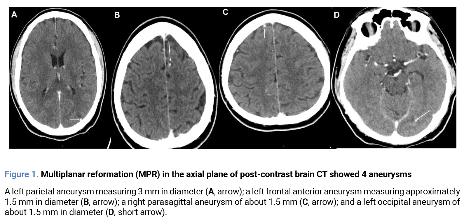
DSA also showed a 1 mm aneurysm of a distal branch of the right occipital artery (Figure 2C, long arrow), not evidenced on brain computed tomography angiography (CTA). This IC-IA, as well as two others (1-1.5 mm) arising from the left callosomarginal artery (Figure 2B, white arrow), was not treated due to the risk of ischemia in the event of occlusion of the main artery.
Follow-up TTE/TEE at HD 3 and 9 showed a stabilization of the lesions. A seven-month follow-up brain-CTA showed the disappearance of all the IIAs. The patient was doing well until 3 years later, when he presented with native MV Streptococcus vestibularis IE.
This case presents three clinical issues. First, the occurrence of multiple IIAs; second, the question of whether or not to treat silent and multiple IAs (and, if so, what type of treatment to use); and third, the A. defectiva IE profile.
IC-IA in IE are multiple in 19% to 33.3% of cases (with a range of two to nine aneurysms per patient). They are incidentally diagnosed in 30% of cases. Endovascular (EV) treatment is the most suitable approach in cases of multiple IC-IAs. In the present case, the aneurysms had all the characteristics of infectious aneurysms: small, distally located, and multiple.
Interestingly, in the present case, we observed that all the IIAs, incidentally diagnosed, arose solely from the territory of the left internal carotid artery (ICA), as both anterior cerebral arteries (ACAs) were supplied by the left ICA. This raises the following question: did these six IIAs develop because of a rain of emboli, which would have migrated via the ICA?.
As in other species, it is plausible that the IAs developed because of the subacute nature of the process; if bacteria have been released into the blood for 2-3 months, after seeding the vasa vasorum, aneurysms may occur.
Given their rarity, no prospective study of the natural course of IC-IAs in IE has been able to provide their rupture rate. However, it is well known that IC-IAs are dynamic lesions with an unpredictable course, that they carry a high risk of rupture, that they can have a rapid onset [10] and rupture rapidly, and that they have the potential for rapid growth and rupture [11].
The management of unruptured IC aneurysms and of IC-IAs is still discussed [12]. The first literature review about the long-term outcome of IC-IAs [13] showed a failure rate of antibiotic therapy alone of 40%, with a mortality of 24%. The systematic review by Ragulojan [14] about EV, surgical, and antibiotic treatment of IC-IAs showed an increasing trend of EV treatment, and in the context of IE, a strong preference for EV treatment before cardiac surgery. In our institution, our approach is to opt for EV treatment as soon as possible and before cardiac surgery for all types of germs.
A. defectiva is a fastidious organism that grows more slowly than other streptococci; thus, cultivation and identification are often difficult, and it is necessary to extend the incubation period beyond 5 days.
A. defectiva often involves patients who have undergone dental procedures. Our patient had no history of dentistry, but numerous colonic polyps were found and resected. Historically, A. defectiva IE is associated with a higher rate of morbidity and mortality, embolism, and heart failure than endocarditis caused by other streptococci. MV involvement is the most common, followed by the aortic valve, and more rarely, the tricuspid and pulmonary valves are concerned.
In our single-centre IE cohort (2014-2023, 210 cases), we observed only 2 cases (1%), the same incidence as that previously reported.
To date, eleven patients with A. defectiva IE, including ours, have been reported with concomitant IIAs [1-9]. Three (33%) of the 9 patients with vascular imaging presented with multiple IIAs: 3 IIAs in an adult [8], 3 IIAs in an 8-year-old child [7], and 6 in our patient.
Considering the cardiac background, a native valve was involved in all 11 reported cases. A pre-existing MV disease was present in 5 cases (45.4%) [1,2,5, present case], while a congenital heart disease was present in 23.7% of cases.
This analysis based on these 11 cases reinforces the recommendations of Rhodes et al. [5] based on 2 personal cases and 2 cases from the literature [1,2,5]. Currently, despite the extremely limited number of published cases, we could suggest that, in patients with MV IE due to A. defectiva, IC-IAs should be suspected, and clinicians should consider imaging the central nervous system, in accordance with the recommendations provided in the clinical practice guidelines published in 2023 [15].
Indeed, brain CT or MRA is recommended in patients with IE and suspected infective cerebral aneurysms. If non-invasive techniques are negative and the suspicion of an infectious aneurysm remains, invasive angiography should be considered.
Our case provides several lessons. First, it demonstrates that Abiotrophia defectiva IE can be complicated by IIA, even that these aneurysms may be multiple, which justifies the need for brain imaging in this context. Second, the mitral valve is most often involved, and, moreover, congenital mitral valve lesions, especially mitral valve prolapse, may be present. Finally, EV treatment appears to be a safe option before cardiac surgery.
Funding
No funding was received for the research, authorship, and/or publication of this article.
Author contribution
Conceptualization: M.B. and J.P.L.; Investigation: M.B.; Writing – original draft: M.B.; Supervision: S.L.; Writing – review & editing: M.B. and J.P.L. All authors have read and approved the submitted manuscript.
Conflicts of interest
No commercial, financial, or personal conflicts of interest were identified in relation to the research, authorship, and/or publication of this article.
AI statement
No artificial intelligence tools were used in the writing, analysis, interpretation, or review of this article.
References
- Yang Y-S, Shang S-T, Lin J-C, Chiu C-H, Chang F-Y. A ruptured cerebral mycotic aneurysm caused by Abiotrophia defectiva endocarditis. Am J Med Sci. 2010;339(2):190-1. Available from: https://doi.org/10.1097/MAJ.0b013e3181be6586
- Kohok DD, Parashar A, Punnam V, Tandar A. Subarachnoid hemorrhage in a patient with Abiotrophia defectiva endocarditis. Am J Med Sci. 2011;341(2):157-9. Available from: https://doi.org/10.1097/MAJ.0b013e3181f99cc7
- Flor-de-Lima F, Lisboa L, Sarmento A, et al. Mycotic brain aneurysm and cerebral hemorrhagic stroke: a pediatric case report. Eur J Pediatr. 2013; 172 (9):1285-1286. doi: 10.1007/s00431-013-2032-5
- Rhodes HM, Hirigoyen D, Shabnam L, Williams DN, Hansen GT. Infective endocarditis due to Abiotrophia defectiva and Granulicatella spp. complicated by infectious intracranial cerebral aneurysms: a report of three cases and review of the literature. J Med Microbiol. 2016;65(6):493–9. Available from: https://doi.org/10.1099/jmm.0.000260
- Cheng DT, Kim PM, Young T, Nikolarakos D. Abiotrophia defectiva bacterial endocarditis complicated by an infective intracranial mycotic aneurysm - The need for aggressive odontogenic source control. J Oral Biol Craniofac Res. 2022;12(5):713–5. Available from: https://doi.org/10.1016/j.jobcr.2022.08.021
- Du Y, Zhang Z, Chen C, Xia H, Zhang H, Guo Z, et al. Case report: Report of infective endocarditis caused by Abiotrophia defectiva and literature review. Front Pediatr. 2022;10:894049. Available from: https://doi.org/10.3389/fped.2022.894049
- Mehta N, Baskind M, Collins K, Acostamadiedo JM. Mycotic Aneurysm from Abiotrophia Defectiva Causing Subdural Hematoma with Herniation. Adv Infect Dis. 2022;12(04):715–20. Available from: https://doi.org/10.4236/aid.2022.124050
- Toda E, Kajiya T, Higo K, Ito K, Ueno M, Takaoka J, et al. Association of infective endocarditis caused by Abiotrophia defectiva with mycotic aneurysm formation: A case report and the review of the literature. J Cardiol Cases. 2023;28(4):161–3. Available from: https://doi.org/10.1016/j.jccase.2023.06.002
- Tokunaga S, Miyakoshi A, Ito K, Sato T. Delayed formation and rupture of intracranial aneurysm following Abiotrophia defectiva endocarditis. BMJ Case Rep. 2024;17(1):e258922. Available from: https://doi.org/10. 1136/bcr-2023-258922
- Samaniego EA, Ortega S, Stander L, Leira EC. Development of a cerebral mycotic aneurysm within 5 days. Neurol Clin Pract. 2017;7(5):449–50. Available from: https://doi.org/10.1212/CPJ.0000000000000315
- Boukobza M, Naggara O, Duval X, Laissy J-P. Acute enlargement, morphological changes, and rupture of intracranial infectious aneurysm in infective endocarditis. Serial imaging. J Clin Neurosci. 2020;82(Pt B):237–40. Available from: https://doi.org/10.1016/j.jocn.2020.11.001
- Rinkel GJE, Ruigrok YM, Krings T, Etminan N, Vergouwen MDI. Advances in screening and management of unruptured intracranial aneurysms. Lancet Neurol. 2025;24(11):958–68. Available from: https://doi.org/10.1016/S1474-4422(25)00265-0
- Alawieh AM, Dimisko L, Newman S, Grossberg JA, Cawley CM, Pradilla G, et al. Management and long-term outcomes of patients with infectious intracranial aneurysms. Neurosurgery. 2023;92(3):515–23. Available from: https://doi.org/10.1227/neu.0000000000002235
- Ragulojan R, Grupke S, Fraser JF. Systematic review of endovascular, surgical, and conservative options for Infectious Intracranial aneurysms and cardiac considerations. J Stroke Cerebrovasc Dis. 2019;28(3):838–44. Available from: https://doi.org/10.1016/j.jstrokecerebrovasdis.2018.11.035
- Delgado V, Ajmone Marsan N, de Waha S, Bonaros N, Brida M, Burri H, et al. 2023 ESC Guidelines for the management of endocarditis. Eur Heart J. 2023;44(39):3948–4042. Available from: https://doi.org/10.1093/eurheartj/ehad193
Explore
Articles
Volumes
About the journal
Most read
Recomendaciones para la prevención y el tratamiento de las infecciones respiratorias en adultos mayores institucionalizados
Revista Española de Quimioterapia
Acute infectious gastroenteritis in childhood: the role of rapid multiplex molecular syndromic panels in diagnosis and clinical management
Revista Española de Quimioterapia
Características epidemiológicas de pacientes con aislamientos de Enterobacterales productores de carbapenemasas-NDM en un hospital de alta complejidad
Revista Española de Quimioterapia
Review on the management of intra-abdominal candidiasis
Revista Española de Quimioterapia
Neoscytalidium dimidiatum: a rare locally acquired deep cutaneous infection in a renal transplant recipient
Revista Española de Quimioterapia