Original
April 2025, Volume 38, nº 2
14 min
Post-COVID-19 tuberculosis in southeastern Spain: incidence, risk factors and the role of latent tuberculosis infection screening
José Antonio Peregrina-Rivas1 , Daniel Fernández-Reyes1 , Adolfo de Salazar-González2,3,4 , Fernando García-García2 , Miguel Ángel Montero-Alonso5 , José Hernández-Quero6 , Emilio Guirao-Arrabal*7
1Internal Medicine Unit, Hospital Universitario Clínico San Cecilio, Granada, Spain
2Microbiology Unit, Hospital Universitario Clínico San Cecilio, Granada, Spain
3Instituto de Investigación Ibs, Granada, Spain
4Ciber de Enfermedades Infecciosas, CIBERINFEC, ISCIII, Madrid, Spain
5Department of Statistics and O.I., Faculty of Medicine, University of Granada, Granada, Spain
6Medicine Department, University of Granada, 18016 Granada, Spain
7Infectious Diseases Unit, Hospital Universitario Clínico San Cecilio, Granada, Spain
https://doi.org/10.37201/req/111.2024
Receipt Date:
23/11/2024
Acceptance Date:
15/01/2025
Publication Date:
05/02/2025
How to cite
Peregrina-Rivas JA, Fernández-Reyes D, de Salazar-González A, García-García F, Montero-Alonso MA, Hernández-Quero J, Guirao-Arrabal E. Post-COVID-19 tuberculosis in southeastern Spain: incidence, risk factors and the role of latent tuberculosis infection screening. Rev Esp Quimioter. 2025 Feb 6;38(2):108-114. doi: 10.37201/req/111.2024
Metrics
404
Views
68
Downloads
Share
Share
Abstract (EN)
Introduction: The incidence of coinfections and superinfections following SARS-CoV-2 pneumonia has garnered increasing attention, with complications arising from various pathogens, including viruses, bacteria, and fungi. Nevertheless, the relationship between COVID-19 and tuberculosis (TB) is not fully understood. This study investigates the incidence and risk factors for post-COVID-19 pulmonary TB in a low TB prevalence area in southeastern Spain, alongside the influence of COVID-19 on indeterminate results in the Quantiferon-TB Gold Plus (QFT-Plus) test and its prognostic role.
Material and methods: A retrospective cohort study was conducted involving 475 hospitalized COVID-19 from March 2020 to March 2022, all of them with a QFT-Plus performed.
Results: The study found three cases of pulmonary TB in the post-COVID-19 period, yielding an incidence density of 3.56 cases per 1000 patient-years, all associated with chronic systemic corticosteroid therapy. Notably, the percentage of indeterminate QFT-Plus results during COVID-19 was significantly higher than when it was performed before the disease (16.82% vs. 3.37%). Patients with indeterminate results exhibited elevated inflammatory markers and a greater need for invasive mechanical ventilation, correlating with more severe disease, although without statistical significance.
Conclusions: The findings suggest that prolonged systemic corticosteroid therapy is a common risk factor for pulmonary TB development and that systematic LTBI screening may not be necessary for all COVID-19 patients unless prolonged corticosteroid treatment is foreseen. This study highlights the need for further research to clarify the relationship between COVID-19 and post-COVID-19 pulmonary TB, as well as the prognostic implications of QFT-Plus results.
Keywords (EN):
fármacos inmunosupresores
Tuberculosis
infección tuberculosa latente
QuantiFERON
COVID-19
Introduction
There is a growing interest in the incidence of coinfections and superinfections after SARS-CoV-2 pneumonia requiring hospitalization. Co/superinfection can be produced by other viruses (influenza A and B, respiratory syncytial virus), bacteria (Streptococcus pneumoniae, Staphylococcus aureus, Klebsiella pneumoniae) or fungi (mainly Aspergillus spp.). These complications have been observed in 16-19% of these patients and are associated with increased morbidity and mortality [1,2]. In addition to this, there is a risk for latent infections reactivation such as for tuberculosis (TB). Approximately one-third of the global population is estimated to have latent tuberculosis infection (LTBI) [3], which can be reactivated in situations of decreased lymphocyte function or quantity, such as human immunodeficiency virus (HIV) infection or chronic treatment with immunosuppressive drugs, such as corticosteroids [3-5]. Taking 15 milligrams of prednisone or equivalent daily doses for at least one month is considered to result in an increased risk of LTBI reactivation [6]. Thus, moderate-severe COVID-19, characterized by lymphopenia and treatment with high doses of corticosteroids, sometimes in combination with other immunosuppressive drugs (tocilizumab, sarilumab, baricitinib, etc.) could pose a risk situation for LTBI reactivation [7–9]. In this sense, cases of pulmonary TB coinfection with SARS-CoV-2 have already been described in areas of high TB incidence, although the relationship between both pathologies has not been fully elucidated and there are no recommendations on the need to screen and treat LTBI in those hospitalized for COVID-19 [7]. In areas of low TB prevalence, like our country, interferon γ release assays (IGRA) are the technique of choice for the diagnosis of LTBI, as they are more specific than the tuberculin skin test. In recent years, the use of Quantiferon-TB Gold plus (QFT-Plus), a new generation IGRA that increases the sensitivity of the test, has become in widespread used [10]. However, this test could have limited utility in patients on corticosteroid treatment, in whom a higher false-positive rate has been described [11]; and those affected by COVID-19, in whom a higher percentage of indeterminate results has been reported compared to the general population [12]. On the other hand, it has been observed that patients hospitalized for COVID-19 with an indeterminate result in QFT-Plus have more severe forms of the disease and higher mortality than those with a determined result (positive or negative) [13]. The main objective of this study was to evaluate the incidence and risk factors for post-COVID-19 pulmonary TB in an area of low TB prevalence in southeastern Spain. A secondary objective was to evaluate the influence of COVID-19 on the appearance of indeterminate results in QFT-Plus and their prognostic role.Material and methods
A retrospective cohort study was carried out at the Hospital Universitario Clínico San Cecilio in Granada between March, 2020 and March, 2022. Individuals over 18 years old with a positive polymerase chain reaction test for SARS-CoV-2 on nasopharyngeal swab result with COVID-19 pneumonia in need for hospitalization and a QFT-Plus (Qiagen®) test performed during admission or in the two years prior were included. This test employs two Mycobacterium Tuberculosis (MT) antigen tubes, TB1 and TB2. While TB1 peptides are designed to elicit an interferon γ (IFN-γ) response from helper CD4+ T lymphocytes, TB2 contains a series of additional peptides that allow measuring the response of cytotoxic CD8+ T lymphocytes [9]. Thus, to evaluate the risk of developing post-COVID-19 pulmonary TB, all QFT-Plus from the sample were considered. On the contrary, to analyze the prognostic role of the QFT-Plus result in the evolution of COVID-19 and, in turn, the influence of the disease on the test results, only those tests performed during COVID-19 admission were selected. The follow-up period for each patient was two years, or until death or diagnosis of pulmonary TB. An anonymized database of patients was created. Patient demographic variables, comorbidities, laboratory parameters, immunosuppressive treatment received, clinical course during hospitalization and outcome at two years of follow-up were extracted from the digital history of patients. For continuous variables, the median and interquartile range were calculated, while for qualitative variables, the absolute frequency and percentage were presented. In hypothesis contrasts, a p-value less than 5% was considered significant, performing the Mann-Whitney U test for continuous variables and the Chi square test or Fisher's exact test for qualitative variables. To carry out the statistical analysis, the IBM SPSS Statistics for Windows, Version 25.0. Armonk, NY: IBM Corp. program was used. Ethics: The study was approved by ethics committee of our center (internal code 1280-N-22).Results
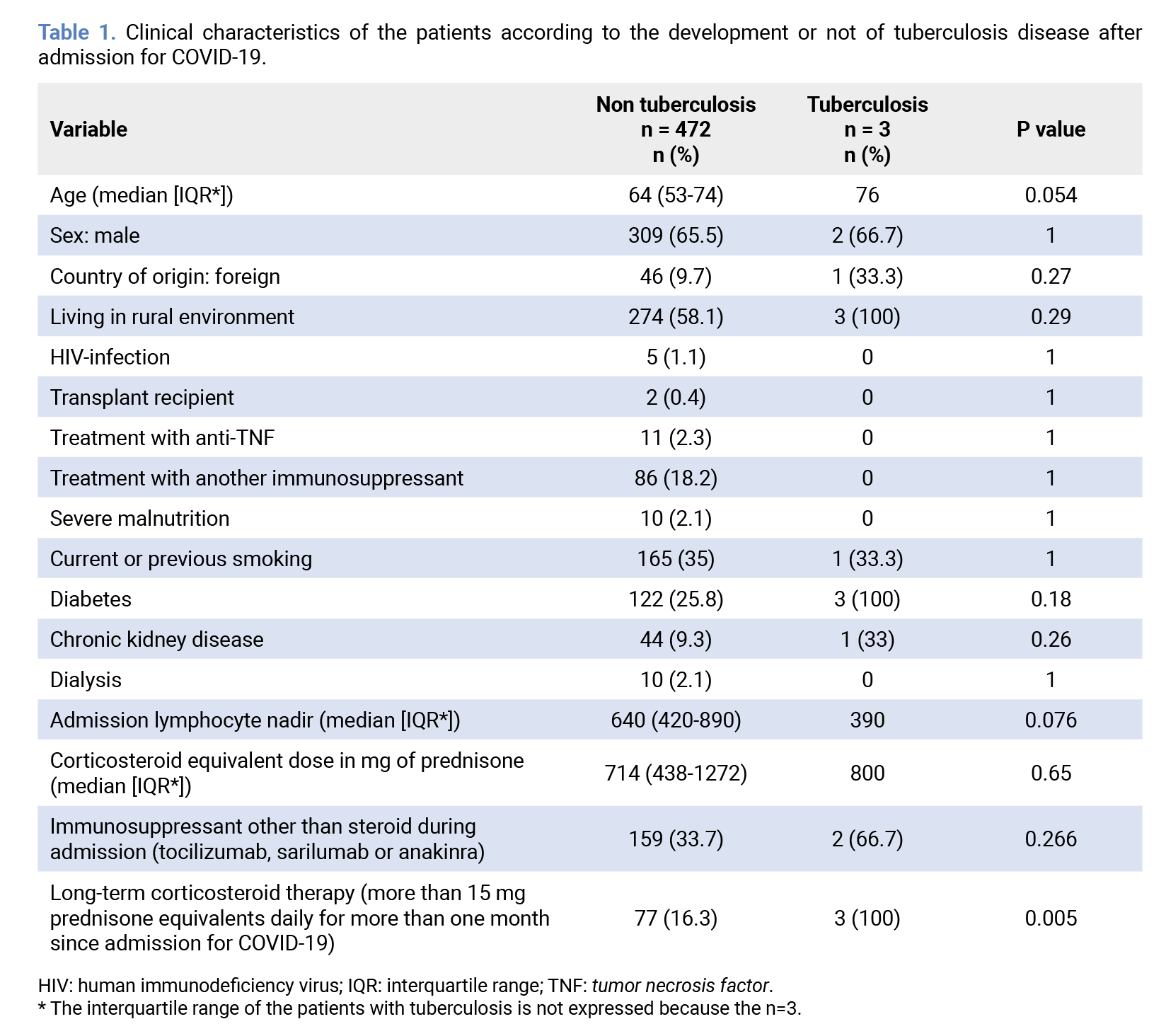
A total of 475 patients were included in our study, as seen in the flowchart (Figure 1). 366 had a negative QFT-Plus (77.05%), 65 had a positive result (13.68%) and the remaining 44 (9.26%) had an indeterminate result. Of the 65 patients with a positive QFT-Plus result, two of them had previously received a complete 6 months antituberculosis treatment course for active pulmonary TB and 21 had received treatment for LTBI: ten with isoniazid for nine months, three wit isoniazid for six months, six with isoniazid and rifampicin for three months and two with a treatment schedule not recorded in the clinical history.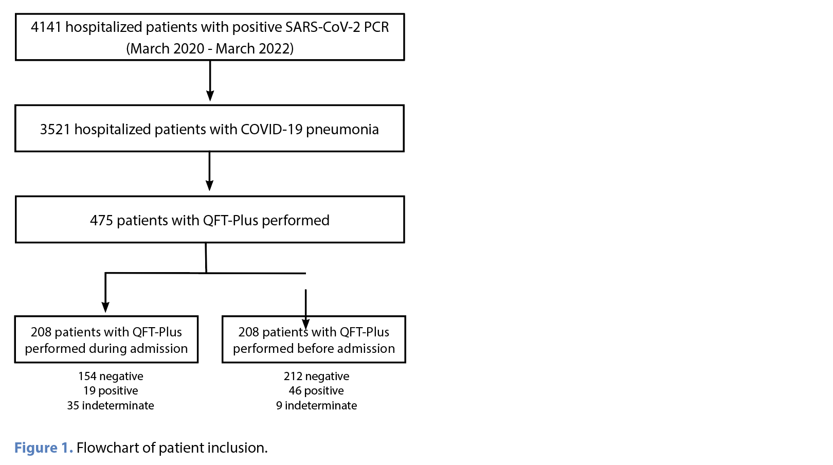 By the end of the follow-up period, three cases of pulmonary TB were diagnosed, yielding an incidence rate of 3.56 cases per 1000 patient-years and a prevalence of 0.63% (3/475). All cases occurred in patients over 70 years old who were receiving chronic systemic corticosteroid therapy for various conditions. One of them was a male patient with a negative QFT-Plus result, who was a smoker affected by chronic obstructive pulmonary disease (COPD) with frequent exacerbations that needed systemic and inhaled corticosteroid therapy. The other two patients were women with positive result in QFT-Plus test, which represented an incidence density of 17.83 cases per 1000 patient-years and a prevalence of 3.17% (2/63) in the subgroup of patients with LTBI. These women had not chronic respiratory pathology and one of them was from Morocco, nevertheless she had resided in Spain for ten years. Despite they were not users of corticosteroid or another immunosuppressant before COVID-19, they were left with chronic systemic corticosteroid therapy after admission: one due to the development of pulmonary fibrosis and the other due to marked asthenia residual to COVID-19. Although the male patient survived, the two females died during follow-up due to pulmonary TB. None of these three cases had previously received treatment for LTBI or TB. Long-term systemic corticosteroid therapy, defined as the administration of 15 milligrams or more of prednisone (or equivalent) doses for over one month following admission for COVID-19, was significantly associated with the development of pulmonary TB. Pulmonary TB cases were older and had lower nadir lymphocyte counts during hospitalization for COVID-19 than those who did not develop pulmonary TB, although without reaching statistical significance (Table 1).
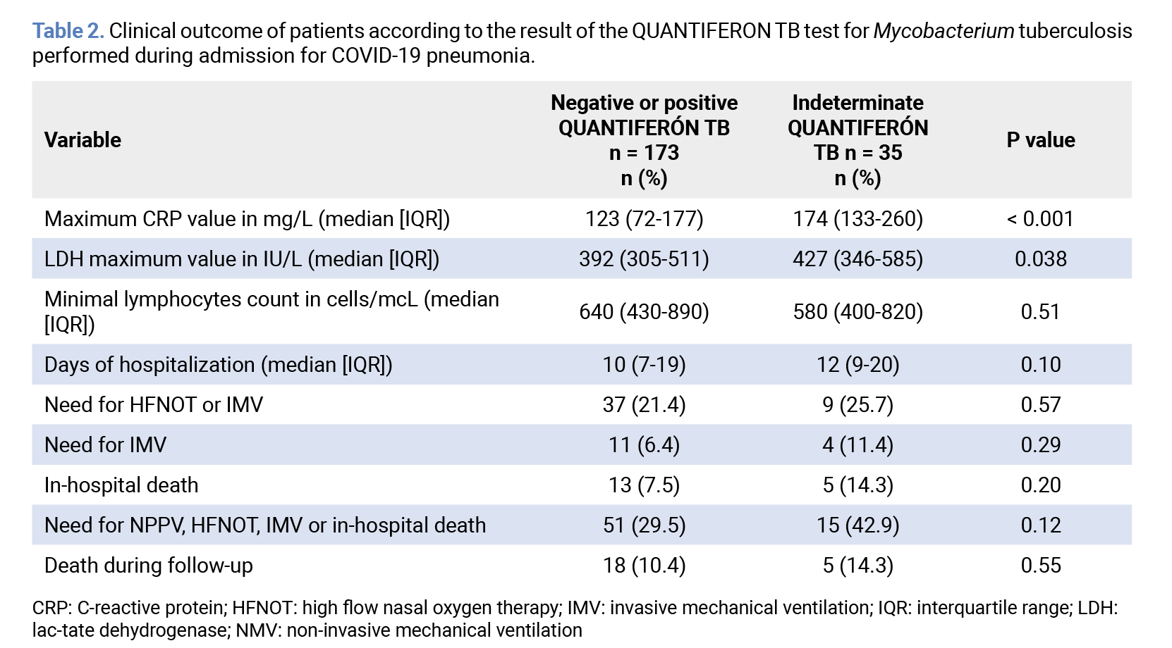
 In order to find out the implication of COVID-19 in QFT-Plus results and the role of this test in COVID-19 prognosis, we analyzed the subgroup of patients in whom QFT-Plus was performed during COVID-19 admission (208 patients) (Table 2). In this way, 16.82% of these tests had an indeterminate result, versus 3.37% when it was performed before COVID-19 (p<0.0005). Patients with an indeterminate result had higher levels of C-reactive protein (CRP) and lactate dehydrogenase (LDH) than those who had a determined result (positive or negative). On the other hand, patients with an indeterminate result had a greater need for high glow nasal oxygen therapy (HFNOT), non-invasive mechanical ventilation (NMV) or invasive mechanical ventilation (IMV), a longer hospital stay and a higher probability of death during admission compared to those who had a positive or negative result, although without reaching statistical significance (Table 2).
Discussion
At the conclusion of follow-up, three patients in our cohort developed post-COVID-19 pulmonary TB: one with a negative QFT-Plus result, and the other two with positive results. Although the patient with a negative result could be assumed as primary TB, the use of systemic corticosteroid therapy is associated with false negative QTF-Plus results [11]. Therefore, it cannot be ruled out that our patient, a habitual user of corticosteroids for his COPD, suffered a reactivation of LTBI instead of a primary TB. Of the two cases observed in patients with positive QFT-Plus, they did not carry out LTBI treatment and both received chronic systemic corticosteroid therapy after COVID-19. We did not find any post-COVID-19 pulmonary TB in those patients receiving standard corticosteroid regimens established in clinical trials, in combination or not with other immunosuppressants [14,15]. In this way, a common risk factor for the development of pulmonary TB in our cohort was prolonged systemic corticosteroid therapy [4]. Another characteristic common to all three cases was being over 70 years of age. In this regard, it is known that aging implies a worse functioning of CD4+ T lymphocytes and a lower production of interleukin 2 and IFN-γ, which implies a deterioration of cellular immunity that predisposes to the reactivation of latent infections such as tuberculosis [16]. This finding suggests that systematic screening for LTBI may not be necessary in all patients hospitalized for COVID-19, except for those who are likely to require prolonged systemic corticosteroid therapy. This could be especially interesting in older subjects with marked lymphopenia during admission for COVID-19, although further studies are needed to establish cut-off points for age and lymphocytes. Another notable fact from our study was that the percentage of indeterminate QFT-Plus results when it was performed during COVID-19 infection was significantly higher than when it was done before suffering from the disease. This could be due to lower production of IFN-ү by lymphocytes of patients with COVID-19, as has been previously reported [17]. A low IFN-ү production leads to greater differentiation of helper lymphocytes (Th0) to Th2 instead of Th1, which causes an exacerbated immune response that contributes to lung damage in COVID-19 and worsen patients’ prognosis [12,18]. This is consistent with our results, in which patients with COVID-19 and an indeterminate QFT-Plus result at admission presented higher inflammatory parameters (CRP and LDH) than those who yielded a positive or negative result in this test. Also, these patients needed greater support with IMV and had higher mortality than those who presented a negative or positive result, although without reaching statistical significance. In this regard, in a study carried out on patients admitted to an Intensive Care Unit before the COVID-19 pandemic, a higher mortality was described for those with an indeterminate QFT-Plus result, compared to those who presented a positive or negative result [19]. This was related to lymphocyte anergy typical of severely ill patients. In subsequent studies with patients affected by COVID-19, higher hospital mortality has also been described for those with indeterminate results during admission, in relation to the defects in cellular immunity that SARS-CoV-2 causes in these patients [13,20]. Taking this into account, it is likely that the lack of statistical significance observed in our study for the prognostic variables was due to an insufficient sample. Our work has several limitations. The main ones are its retrospective nature and its unicentric character, which limit its external validity. Furthermore, the information was obtained from the patients’ medical records, which limits the quality of the data obtained. On the other hand, the number of patients included to analyze the prognostic role of the QFT-Plus result was not high enough to obtain statistically significant results in most of the variables. Further studies are required to better clarify the prognostic value of QFT-Plus in this context. The main strength of our work lies in the fact that it is, as far as we know, the only longitudinal study carried out in our environment to evaluate the risk of post-COVID-19 TB. This is of interest not only in COVID-19, since the use of immunosuppressants for short periods of time is a common therapeutic resource in other pathologies (pneumonias produced by microorganisms other than SARS-CoV-2, hemophagocytic syndrome, etc.). This study highlights the need to consider the risk of TB following prolonged corticosteroid courses, even in countries with a low TB incidence. In conclusion, the three cases of pulmonary TB observed in our study corresponded to elderly patients who received prolonged corticosteroid courses after being hospitalized for COVID-19, exceeding the corticosteroid therapy regimens established for the treatment of this disease in clinical trials. Our proposal is to perform LTBI screening and treatment in those patients over seventy years who are hospitalized for COVID-19 in whom prolonged treatment with corticosteroids can be foreseen [21]. In addition, the high percentage of indeterminate results in QFT-Plus questions the usefulness of systematic LTBI screening in patients with COVID-19. On the other hand, those admitted with an indeterminate result presented analytical data compatible with more severe forms of COVID-19, which has been related in previous studies to a worse prognosis. Further studies are necessary to clarify the relationship between COVID-19 and post-COVID-19 pulmonary TB, as well as the prognostic implication of QFT-Plus results. Acknowledgements We thank all the health professionals who worked tirelessly to combat COVID-19. Funding None to declare. Conflicts of interest The authors declare no conflicts of interest. Author contributions Conceptualization, JA P-R. and E G-A; methodology, MA M-A; software, MA M-A; validation, D F-R, A S-G and E G-A; formal analysis, MA M-A and D F-R; investigation, JA P-R; resources, F G-G and A S-G; data curation, JA P-R; writing—original draft preparation, JA P-R; writing—review and editing, D F-R, A S-G and E G-A; visualization, J H-Q and E G-A; supervision, J H-Q; project administration, J H-Q. All authors have read and agreed to the published version of the manuscript.References
- Kurra N, Woodard PI, Gandrakota N, Gandhi H, Polisetty SR, Ang SP, et al. Opportunistic Infections in COVID-19: A Systematic Review and Meta-Analysis. Cureus 2022 31;doi: 10.7759/cureus.23687.
- Musuuza JS, Watson L, Parmasad V, Putman-Buehler N, Christensen L, Safdar N. Prevalence and outcomes of co-infection and superinfection with SARS-CoV-2 and other pathogens: A systematic review and meta-analysis. PLoS ONE 2021 6;16:e0251170. doi: 10.1371/journal.pone.0251170.
- Shariq M, Sheikh JA, Quadir N, Sharma N, Hasnain SE, Ehtesham NZ. COVID-19 and tuberculosis: the double whammy of respiratory pathogens. Eur Respir Rev 2022 30;31:210264. doi: 10.1183/16000617.0264-2021.
- Ai J-W, Ruan Q-L, Liu Q-H, Zhang W-H. Updates on the risk factors for latent tuberculosis reactivation and their managements. Emerging Microbes & Infections 2016;5:1–8. doi: 10.1038/emi.2016.10.
- Machuca I, Vidal E, De La Torre-Cisneros J, Rivero-Román A. Tuberculosis en pacientes inmunodeprimidos. Enfermedades Infecciosas y Microbiología Clínica 2018;36:366–74. doi: 10.1016/j.eimc.2017.10.009.
- Jick SS, Lieberman ES, Rahman MU, Choi HK. Glucocorticoid use, other associated factors, and the risk of tuberculosis. Arthritis Rheum 2006 15;55:19–26. doi: 10.1002/art.21705.
- Siranart N, Sowalertrat W, Sukonpatip M, Suwanpimolkul G, Torvorapanit P. First case series and literature review of coronavirus disease 2019 (COVID-19) associated pulmonary tuberculosis in Southeast Asia: Challenges and opportunities. Journal of Infection and Public Health 2023;16:80–9. doi: 10.1016/j.jiph.2022.11.029.
- Visca D, Ong CWM, Tiberi S, Centis R, D’Ambrosio L, Chen B, et al. Tuberculosis and COVID-19 interaction: A review of biological, clinical and public health effects. Pulmonology 2021;27:151–65. doi: 10.1016/j.pulmoe.2020.12.012.
- Yang H, Lu S. COVID-19 and tuberculosis. Journal of Translational Internal Medicine 2020 1;8:59–65. doi: 10.2478/jtim-2020-0010.
- Pai M, Behr M. Latent Mycobacterium tuberculosis Infection and Interferon-Gamma Release Assays. Microbiol Spectr 2016 14;4:4.5.24. doi: 10.1128/microbiolspec.TBTB2-0023-2016.
- Bartalesi F, Vicidomini S, Goletti D, Fiorelli C, Fiori G, Melchiorre D, et al. QuantiFERON-TB Gold and the TST are both useful for latent tuberculosis infection screening in autoimmune diseases. European Respiratory Journal 2009 1;33:586–93. doi: 10.1183/09031936.00107608.
- Ward JD, Cornaby C, Schmitz JL. Indeterminate QuantiFERON Gold Plus Results Reveal Deficient Interferon Gamma Responses in Severely Ill COVID-19 Patients. J Clin Microbiol 2021 20;59:e00811-21. doi: 10.1128/JCM.00811-21.
- Solanich X, Fernández-Huerta M, Basaez C, Antolí A, Rocamora-Blanch G, Corbella X, et al. Clinical Significance of Indeterminate QuantiFERON-TB Gold Plus Assay Results in Hospitalized COVID-19 Patients with Severe Hyperinflammatory Syndrome. JCM 2021 26;10:918. doi: 10.3390/jcm10050918.
- The RECOVERY Collaborative Group. Dexamethasone in Hospitalized Patients with Covid-19. N Engl J Med 2021 25;384:693–704. doi: 10.1056/NEJMoa2021436.
- Abani O, Abbas A, Abbas F, Abbas M, Abbasi S, Abbass H, et al. Tocilizumab in patients admitted to hospital with COVID-19 (RECOVERY): a randomised, controlled, open-label, platform trial. The Lancet 2021;397:1637–45. doi: 10.1016/S0140-6736(21)00676-0.
- Schaaf HS, Collins A, Bekker A, Davies PDO. Tuberculosis at extremes of age. Respirology 2010;15:747–63. doi: 10.1111/j.1440-1843.2010.01784.x.
- Palacios-Gutiérrez J-J, Rodríguez-Guardado A, Arias-Guillén M, Alonso-Arias R, Palacios-Penedo S, García-García J-M, et al. Clinical and Epidemiological Correlates of Low IFN-Gamma Responses in Mitogen Tube of QuantiFERON Assay in Tuberculosis Infection Screening During the COVID-19 Pandemic: A Population-Based Marker of COVID-19 Mortality? Archivos de Bronconeumología 2022;58:649–59. doi: 10.1016/j.arbres.2022.01.011.
- Huang C, Wang Y, Li X, Ren L, Zhao J, Hu Y, et al. Clinical features of patients infected with 2019 novel coronavirus in Wuhan, China. The Lancet 2020;395:497–506. doi: 10.1016/S0140-6736(20)30183-5.
- Huang C-T, Ruan S-Y, Tsai Y-J, Kuo P-H, Ku S-C, Lee P-L, et al. Effects of acute critical illnesses on the performance of interferon-gamma release assay. Sci Rep 2016 25;6:19972. doi: 10.1038/srep19972.
- Remy KE, Mazer M, Striker DA, Ellebedy AH, Walton AH, Unsinger J, et al. Severe immunosuppression and not a cytokine storm characterizes COVID-19 infections. JCI Insight 2020 3;5:e140329. doi: 10.1172/jci.insight.140329.
- Youssef J, Novosad SA, Winthrop KL. Infection Risk and Safety of Corticosteroid Use. Rheumatic Disease Clinics of North America 2016;42:157–76. doi: 10.1016/j.rdc.2015.08.004.
Explore
Articles
Volumes
About the journal
Most read
Recomendaciones para la prevención y el tratamiento de las infecciones respiratorias en adultos mayores institucionalizados
Revista Española de Quimioterapia
Acute infectious gastroenteritis in childhood: the role of rapid multiplex molecular syndromic panels in diagnosis and clinical management
Revista Española de Quimioterapia
Características epidemiológicas de pacientes con aislamientos de Enterobacterales productores de carbapenemasas-NDM en un hospital de alta complejidad
Revista Española de Quimioterapia
Review on the management of intra-abdominal candidiasis
Revista Española de Quimioterapia
Neoscytalidium dimidiatum: a rare locally acquired deep cutaneous infection in a renal transplant recipient
Revista Española de Quimioterapia