Review
December 2025, Volume 38, nº 6
50 min
Review on the management of intra-abdominal candidiasis
Pedro Puerta-Alcalde1,10,11 , Jesús Guinea2,3,4,5,* , Emilio Maseda6,7, Rafael Zaragoza8,9
1Hospital Germans Trias i Pujol, Badalona, Spain
2Clinical Microbiology and Infectious Diseases, Hospital General Universitario Gregorio Marañón, Madrid, Spain
3Instituto de Investigación Sanitaria Gregorio Marañón, Madrid, Spain
4CIBER Enfermedades Respiratorias-CIBERES (CB06/06/0058), Madrid, Spain
5Faculty of Health Sciences, HM Hospitals, Universidad Camilo José Cela, Madrid, Spain
6Anesthesia and Reanimation Service, Hospital Quirónsalud Valle del Henares, Torrejón de Ardoz, Madrid, Spain
7Department of Pharmacology and Toxicology, Complutense University of Madrid, Madrid, Spain
8Intensive Care Unit, Hospital Universitario Dr. Peset, Valencia, Spain
9Fundación Micellium, Valencia, Spain
10Fundació Lluita contra les Infeccions, Badalona, Spain
11Universitat de Vic (UVic) - Universitat Central de Catalunya, Vic, Spain
http://www.doi.org/10.37201/req/095.2025
Receipt Date:
09/09/2025
Acceptance Date:
27/09/2025
Publication Date:
20/10/2025
How to cite
Puerta-Alcalde P, Guinea J, Maseda E, Zaragoza R. Review on the management of intra-abdominal candidiasis. Rev Esp Quimioter. 2025; 38(6):454-470. Available from: http://www.doi.org/10.37201/req/095.2025
Metrics
1514
Views
264
Downloads
Share
Share
Abstract (EN)
Intra-abdominal candidiasis, which includes Candida peritonitis and Candida produced intra-abdominal abscesses, accounts for 10-30% of all intra-abdominal infections diagnosed in the intensive care units. Intra-abdominal candidiasis associates to longer hospital stay, and significantly higher morbidity and mortality. Although the management of invasive candidiasis has greatly improved in these past years, the optimal management of intra-abdominal candidiasis remains elusive. Questions concerning the microbiological diagnosis, optimal antifungal drugs doses, diffusion through peritoneal fluid, and the value of liposomal amphotericin B as first-line treatment, remain unanswered. In this article, three important issues concerning intra-abdominal candidiasis have been re-viewed: microbiological diagnosis and risk of antifungal resistance emergence, pharmacokinetic/pharmacodynamic particularities of antifungals, and clinical management on the daily practice. Only an optimized multidisciplinary approach combining rapid diagnostics, tailored antifungal therapy, and effective source control will improve the management and prognosis of patients with intra-abdominal candidiasis.
Keywords (EN):
Invasive fungal infection
Invasive candidiasis
Echinocandins
Intra-abdominal candidiasis
Liposomal amphotericin B
1. Introduction
Intra-abdominal candidiasis (IAC), which is the most common type of deep-seated candidiasis, encompasses Candida peritonitis and Candida-produced intra-abdominal abscess [1]. The burden of IAC is remarkable in developed countries. A recent study, which assessed the prevalence of Candida peritonitis in 29 countries, reported an overall average incidence of 1.15 cases per 100,000 inhabitants [2]. Remarkably, the average incidence of Candida peritonitis in Spain was 1.42 cases per 100,000 inhabitants, the fourth-highest incidence reported in the above-mentioned study [2]. Intra-abdominal infections accounts for the second most frequently acquired infections in the intensive care units (ICU), with Candida being responsible for up to 10-30% of cases [3]. IAC is also associated with longer hospital stay, and significantly higher morbidity and mortality rates [4-6]. The management of invasive candidiasis has been remarkably improved in the past few years, mainly due to the launch of new antifungal drugs and the development of clinical guidelines [7,8]. Moreover, standardized definitions for candidemia and deep-seated candidiasis in patients admitted to the ICU have been recently proposed by a multidisciplinary panel of experts, which will certainly help optimize the quality of care and the outcome of patients (criteria for proven and probable deep-seated candidiasis infection can be reviewed in Table 1) [9]. Despite these improvements, several questions concerning the diagnosis of IAC remain unanswered: microbiological tools to diagnose the infection, and the optimal antifungal drugs (and doses) to maximize the drug concentrations in the peritoneal fluid and increase the chances for a better outcome [10]. Furthermore, the specific patient populations at highest risk for IAC who may benefit from early antifungal treatment have yet to be identified.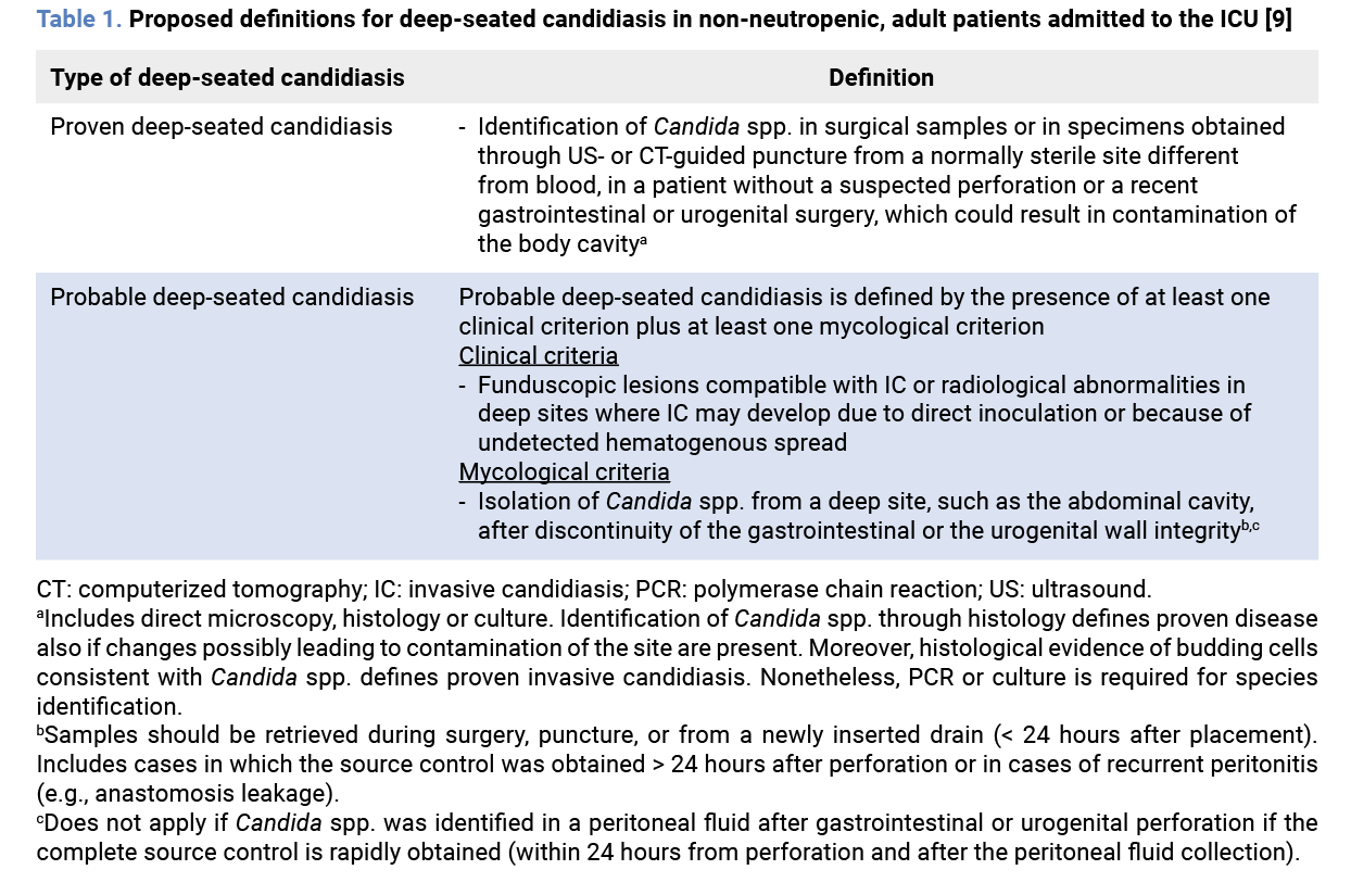 Three relevant topics concerning IAC are here reviewed: the microbiological diagnosis of the infection and antifungal resistance detection, PK/PD particularities of antifungals in this setting, and the clinical management of IAC.
2. Microbiological diagnosis and antifungal resistance detection in IAC
2.1. What is the standard method for the microbiological diagnosis of IAC and their main limitations? The diagnosis of IAC might be challenging in the daily practice due to the non-specific clinical presentation of the infection and the limitations of the currently available diagnostic tests (diagnostic tests currently used in the clinical practice are shown in Table 2) [1,11].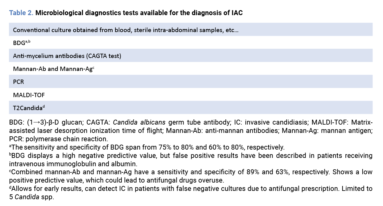 Blood cultures are still the gold standard for the diagnosis of invasive candidiasis. IAC is commonly a consequence of Candida access into the abdominal cavity following gastrointestinal tract disruption; of note, only 5 to 20% of cases will develop secondary candidemia [12]. In a prospective, multicenter study performed in 101 French ICUs, which included a total of 271 eligible adult patients with proven invasive candidiasis, candidemia was found in 184 patients (67.9%), whereas the remaining 87 (32.1%) patients had invasive candidiasis without candidemia [13]. Interestingly, in 70 patients without candidemia, the positive culture was obtained from abdominal specimens at the moment of surgery [13]. In addition to the high frequency of negative blood cultures, the time required for the detection and identification of Candida frequently exceeds 72 hours, significantly delaying the initiation of appropriate antifungal treatment [14]. Accordingly, a study conducted by Nunes et al. pointed out at a time to positivity as the only independent predictor of increased mortality [15]. Similarly, other studies also highlighted that delaying empiric treatment of invasive candidiasis is associated to a higher mortality, especially in patients admitted to the ICU [16,17].
Matrix-assisted laser desorption/ionization time-of-flight mass spectrometry (MALDI-TOF MS) and multiplex polymerase chain reaction (PCR) are new microbiological tools which reduce the time from blood culture detection to final identification of Candida spp. [18]. MALDI-TOF MS requires only 10 to 15 min to identify the Candida spp. and might also be used for antifungal susceptibility tests [18]. PCR performed on blood samples have the highest sensitivity and specificity (90-95% and 90-92%, respectively), reduces the time to diagnosis, and allows Candida spp. identification in many species [18]. Unfortunately, these procedures are only useful once the blood culture has turned positive. New microbiological diagnostic tests are needed to increase the sensitivity of blood cultures, allow faster species identification, optimize the use of antifungal drugs, and help monitor the response to treatment. Despite these interesting results, this tool is no longer available.
2.2. What is the value of non-culture-based diagnostic methods, such as β-D-glucan or Candida PCR, for the diagnosis of IAC? What are their predictive values?
Several meta-analyses have addressed the value of (1→3)-β-D glucan (BDG) for the diagnosis of invasive fungal infection (IFI) [19-21]. For example, a meta-analysis that included a total of 2,979 patients (594 with proven or probable IFIs according to the European Organization for Research and Treatment of Cancer/Mycoses Study Group criteria, and additional 2,385 patients without IFIs) from 16 eligible studies, reported that BDG had a pooled sensitivity of 76.8% (95% confidence interval [CI], 67.1%-84.3%), and a specificity of 85.3% (95% CI, 79.6%-89.7%) [19], with an area under the summary receiver operating characteristic (ROC) curve of 0.89 for the diagnosis of IFI. Nevertheless, the sensitivity of BDG testing to specifically detect proven or probable IC lowered to 75%. A different meta-analysis, which performed a subgroup analysis on the diagnostic accuracy of BDG in patients diagnosed with invasive candidiasis, and included 19 studies, described a sensitivity of 81% (95% CI, 77%-85%), a specificity of 81% (95% CI, 80%-83%), and a ROC curve of 0.90 (95% CI, 0.85-0.95) [20]. However, these data were obtained from patients mainly diagnosed with candidemia. Additionally, BDG concerns stem mainly from false positive results commonly observed due to different reasons such as fungi colonization, some bacteria, certain β-lactams, enteral nutrition, and others. Recent studies have evaluated the role of BDG specifically in patients with IAC [22].
Dupont et al. conducted a prospective, single-center study evaluating serum and peritoneal BDG among patients admitted to the ICU with complicated intra-abdominal infections, and positive or negative fungal cultures [23]. Although there was a trend for higher BDG levels in patients with positive fungal cultures, results were not significant, and the authors concluded that BDG was not useful in the diagnosis or follow-up of these patients. However, many patients had received antifungals prior to BDG processing potentially causing a false negative result of the culture, leading to the misclassification of the positive BDG as a false positive.
On the other hand, a French study recently evaluated the role of BDG in serum and peritoneal samples in 199 patients admitted to the ICU after abdominal surgery for abdominal sepsis, with 87/199 (44%) patients suffering IAC. In this study, both serum and peritoneal BDG were significantly associated with IAC diagnosis, and combining a peritonitis score <3, a serum BDG <3.3 pg/mL, and a peritoneal BDG <45 pg/mL (both using the Wako® test), resulted into 100% negative predictive values (NPV) [24]. In order to increase the BDG testing performance, several authors have proposed the combined use of different tests.
A study that included 434 patients admitted in the ICU with abdominal surgery or acute pancreatitis concluded that BDG antigenemia was superior to Candida score and colonization. Additionally, BDG values decreased when responding to treatment and increased in nonresponse [25]. A BDG value above 259 pg/mL alongside a positive test for Candida albicans germ tube antibody (CAGTA) accurately differentiated Candida colonization from invasive candidiasis in 176 critically ill patients diagnosed with a severe intra- abdominal condition, with a sensitivity of 90.3% [26].
Moreover, the combined use of the tests showed a NPV of 93.9% as long as BDG value was below 259 pg/mL and the CAGTA resulted negative [26]. Other studies have confirmed previous findings, reporting a sensitivity and a NPV superior to 90% when BDG and CAGTA were used in combination [12,27]. This could help clinicians detect patients who might benefit from starting empirical treatment or those who will not [27]. In another study, Xie et al. evaluated the use of a Candida PCR targeting the ITS region in peritoneal fluid samples from ICU surgical patients at high risk of IAC [28]. The assay showed sensitivity, specificity, PPV, and NPV values of 64.7%, 89.4%, 90.8%, and 61.1%, respectively. Combining Candida PCR with BDG increased PPV but decreased sensitivity. Importantly, DNA amplification and culture results were concordant in all but one case of mixed Candida infection.
Among all molecular methods, T2Candida (T2 Biosystems, Lexington, Massachusetts) stands out. It is an innovative nanodiagnostic panel that uses T2 magnetic resonance (T2MR) combined with PCR amplification to detect Candida directly in whole blood samples [29]. The panel is able to detect up to 5 Candida species (C. albicans, C glabrata, C. parapsilosis, C. tropicalis, and C. krusei) and does not require the yeasts to be viable for culture. Some studies have shown good sensitivity values around 90% [29],
and faster mean time to detection compared to blood culture and species identification [30]. Moreover, the mean time to receiving targeted antimicrobial therapy and to empirical therapy de-escalation were significantly faster with T2MR. Likewise, the mean length of ICU admission and the mean length of hospital stay was shorter with T2MR. However, T2MR was not superior to blood culture in terms of allocating patients at the highest risk of mortality [30].
Altogether, available data suggest that T2MR might eventually improve diagnostic accuracy and be used in combination with other microbiological techniques, thus limiting the use of inappropriate treatment and the risk of resistance emergence to antifungal drugs [31]. Nevertheless, some limitations are worthy mentioned, such as the incapability to detect concomitant bacteremia, the limited number of Candida species detected, the impossibility to provide antifungal susceptibility data, its high cost, and limited data from patients with IAC [31]. A recent study compared the use of T2Candida and BDG for the diagnosis of IAC in 134 patients admitted to the ICU or high-dependency unit due to gastrointestinal or necrotizing pancreatitis [32]. Thirteen (10%) patients were diagnosed with IAC with only two of them (15%) presenting concurrent candidemia. T2Candida had lower sensitivity than BDG (36% vs. 82%) but showed excellent specificity and negative predictive value (97% and 94%, respectively). A similar study evaluated T2Candida in 48 high-risk ICU patients with proven IAC in 18 (37.5%) of them [33], with only 2 patients having also candidemia. T2Candida sensitivity/specificity and positive/negative predictive values were 33%/93% and 71%/74%, respectively. Interestingly, IAC was present in 100% of cases with concordant positive T2Candida/BDG and absent in 90% of concordant negative results.
2.3 What are the causative species most frequently implicated in IAC? Are Candida polyfungal infections common?
Several recent studies have assessed the trend of Candida spp. isolated in patients diagnosed with invasive candidiasis. A Spanish tertiary care center studied 166 incident yeast isolates causing fungemia in patients admitted from January 2020 to December 2022, and compared the epidemiology with the one found in two previous periods (2007-2013 and 2014-2019, respectively) [34]. C. albicans was the most frequent isolated specie, although the proportion of isolates started to recede in favor of C. tropicalis and C. parapsilosis. Only in 3 patients (1.8%), two Candida spp. were concomitantly isolated. Antifungal resistance remained low, showing a stable fluconazole resistance rate, an extremely low echinocandin resistance rate, and no resistance to L-AmB [34]. Another prospective study assessed Candida spp. isolates from the bloodstream and the intra-abdominal cavity from patients admitted to 16 Spanish hospitals located in Madrid, Spain, between 2019 and 2021 [35]. Overall, 2,107 Candida isolates from 1,895 patients were analyzed, including blood cultures (n = 1089 [51.7%]) and intra-abdominal samples (n = 1018, [48.3%]).
C. albicans was the most frequent species in both sample types. While C. parapsilosis was the second most common cause of fungemia, C. glabrata complex was the second most frequent species in intra-abdominal samples, accounting for nearly a quarter of positive cases [35]. Interestingly, 130 patients (6.9%) yielded ≥ 2 different species simultaneously isolated from samples collected in the intra-abdominal cavity (n = 104 [5.5%]) or the bloodstream (n = 26 [1.4%]) [35].
2.4 Which is the incidence of infections caused by azole- and/or echinocandin-resistant isolates? Could IAC be a hidden reservoir of strains resistant to antifungals?
Due to the increasing numbers of infections caused by fluconazole-resistant and echinocandin-resistant Candida isolates, the interest on identifying unrecognized niches that could favour resistance to antifungal drugs has grown. For example, in the previously mentioned study [35], fluconazole resistance in C. glabrata isolates was observed in 5.4% of fungemia cases, whereas this percentage nearly doubled to 9.8% in intra-abdominal isolates. Notably, no resistance to L-AmB was reported. Another retrospective study, which included 1,103 samples from 507 patients, described the antifungal resistance profile according to different anatomical compartments: bloodstream (n = 152), normally sterile sites, such as the abdominal cavity, deep organs and deep-seated soft tissues (n = 288), and nonsterile sites, such as the skin and mucosa, the lower respiratory tract and the urine (n = 663) [36]. C. albicans was the most frequent isolate, regardless of the anatomical compartment (63%), while C. glabrata (26.9%) was the second most frequently isolated species in the samples retrieved from the abdominal cavity. A total of 18 patients (3.6%) were diagnosed with fluconazole- resistant (2.2%) or echinocandin-resistant (1.8%) isolates. No resistance to L-AmB was detected. Most patients with fluconazole-resistant and echinocandin- resistant isolates had been previously treated with azoles (63%) and echinocandins (89%), respectively. Fluconazole and echinocandin resistance rates were more frequent in samples collected from the abdominal cavity than from the other studied compartments (3.2% and 3.2%, respectively) [36].
Moreover, the isolates of C. glabrata collected from the abdominal cavity also tended to show a higher rate of fluconazole resistance (11.9% vs 3.2%) and echinocandin resistance (7.1% vs 3.2%) than the isolates of C. glabrata retrieved from the blood cultures. Compartmentalization of antifungal resistance was detected in 6 of 15 patients diagnosed with invasive candidiasis. Except from one patient, the remaining five had susceptible isolates in blood cultures, whereas the resistant isolates originated mostly from the abdominal cavity. The authors concluded that antifungal resistance was mainly associated with Candida glabrata isolates collected from the abdominal cavity, that resistant isolates were most frequently detected in patients with prior antifungal treatment, that some patients exhibited resistance compartmentalization, and that resistance could be overlooked if testing was performed solely on bloodstream isolates. [36]. A more recent study, which included 308 intra-abdominal isolates from 112 patients treated at 7 hospitals located in Madrid from 2019 to 2022, tested for antifungal drug resistance in the initial and sequential isolates from the same species [37].
Overall, fluconazole resistance was detected in 15 of 112 patients (13.4%) and echinocandin resistance in 10 of 112 (8.9%) patients, respectively. No resistance to L-AmB was observed. Resistance would have been overlooked in 11 of 18 patients (61.1%) if only incident isolates had been studied, and it was mainly associated with echinocandin-resistant Candida glabrata. Approximately, 26.7% and 80% of patients with fluconazole or echinocandin-resistant isolates had received or were receiving fluconazole or echinocandins, respectively. The authors concluded that the abdominal cavity could be a reservoir of antifungal resistance, especially echinocandin-resistant C. glabrata, and suggested that testing only incident isolates could have led to underestimating echinocandin resistance in a significant number of patients [37]. In line with this, an earlier study by Shields et al. found echinocandin failure in 13 of 25 (52%) patients with intraabdominal candidiasis [38]. Notably, 24% (6/25) of patients were infected with FKS mutant Candida, a finding that was significantly more frequent in those with echinocandin breakthrough infections (45% vs 6%, p=0.03), reinforcing the concept of a hidden reservoir for resistance development.
3. PK/PD particularities of the antifungal treatment when treating patients diagnosed with IAC
Pharmacokinetics (PK) refers to the processes that affect a drug administered to a patient and encompasses four phases: absorption of a drug from the site of administration, distribution throughout the body, metabolism, and excretion. In turn influence its concentration at the targeted site of action. These processes ultimately condition both the therapeutic and the adverse effects observed in the patient [39]. In contrast, pharmacodynamics (PD) refers to the interaction between the antifungal agent and the fungal organisms, and that interaction dictates the in vitro profile of activity of the drug in question. In the clinical scenario, the interaction among the patient, the drug and the microorganism can be studied by means of the PK/PD interaction and such relationship can be affected when changes on PK, PD, or both occur. Differences in patient-specific factors (e.g. age, severity of the disease, and interaction between drugs), or loss of antifungal susceptibility in isolates causing infections can lead to unpredictable changes in PK/PD parameters, and account for much of the interindividual discrepancies in drug response. Critical ill patients usually show expanded cardiac output, leaky capillaries, altered protein binding, and renal and hepatic dysfunction, which affects the clearance and the volume of distribution [40]. As such, critically ill patients usually show significant changes on the drug’s PK/PD parameters that can greatly affect optimal pharmacotherapy and, ultimately, the patient’s outcome [39,40]. The characteristics of the most common antifungal drugs used in the clinical practice are shown in Table 3.
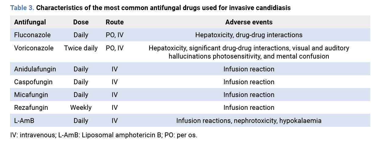 In summary, three main factors contribute to the high risk of suboptimal antifungal concentrations in patients with IAC. First, hydrophilic antifungals, such as echinocandins, are significantly affected by increased volume of distribution (Vd) and enhanced renal clearance, both situations commonly occurring in post-surgical and ICU patients. Second, molecular weight and protein binding further influence drug concentrations and the likelihood of achieving therapeutic targets. Finally, many antifungals exhibit poor penetration into intra-abdominal sites, limiting their effectiveness in deep-seated infections, and posing a risk for potential development of antifungal resistance. A prospective, multinational study performed in 68 ICUs across Europe, which included critically ill patients treated with fluconazole (n = 15), anidulafungin (n = 9), and caspofungin (n = 7), assessed the PK/PD indexes of these drugs at 3 different time points: 30 minutes after completing the intravenous infusion, halfway through the dosing interval, and at the end of the dosing interval [41].
ICU-admitted patients showed greater interindividual variability and lower drug exposures for all three antifungals compared to non-critically ill patients and healthy volunteers. Notably, 33% of patients receiving fluconazole failed to attain the PK/PD target required for optimal outcome [41]. A study conducted in Belgium, in which L-AmB exposure and PK parameters were assessed and compared in two cohorts of critically ill patients (n = 31) and non-critically ill patients (including hematological disease patients and healthy volunteers), described a considerable intra- and intersubject variability in L-AmB [42]. No covariates explaining this variability were identified, including patient-related characteristics [42].
The body weight and the body mass index can influence the PK/PD parameters of antifungal drugs. Interestingly, an analysis of population-based studies concluded that, by 2025, approximately 1 out of 5 individuals will be obese and 1 out of 15 will be diagnosed with severe obesity [43]. An observational PK study conducted at an Australian tertiary referral ICU, which included adult non-obese (n = 11), obese (n = 6) and morbidly obese patients (n = 4), receiving fluconazole either as prophylaxis or targeted treatment for Candida spp. infections, concluded that the standard fluconazole dose (200 mg daily) was insufficient to treat susceptible C. albicans and C. tropicalis isolates in all three groups of patients [44].
The authors suggested that a weight-based loading dose of 12 mg/kg followed by a daily maintenance dose of 6 mg/kg, adjusted by renal function, was a better approach to optimize treatment with fluconazole in obese and morbidly obese patients [44]. A PK study conducted in 11 morbidly obese critically ill adult patients, 10 non-obese critically ill patients, and 10 obese non-critically ill patients who received micafungin for invasive candidiasis (IC) reported inadequate micafungin exposure with the standard 100 mg/24 hours dose, regardless of the Candida species or the patient’s weight. [45]. The authors recommended increasing the dose of micafungin to 150 mg/24 hours to treat C. albicans infections in patients weighing up to 115 kg and 200 mg/24 hours for those surpassing this weight. In the case of infections produced by C. glabrata, a dose of 200 mg/24 hours was recommended for patients weighing up to 115 kg [45]. A different study that determined the PK parameters of anidulafungin in 12 normal-weight subjects and 8 obese subjects concluded that body weight influenced both the clearance and the volume of distribution of the drug, and that a 25% increase in both the loading and maintenance doses could be considered in patients weighing more than 140 kg [46]. Similar findings were also described in the case of caspofungin, suggesting that doses higher than 70 mg/24 hours could be needed to reach PK/PD targets in morbidly obese patients admitted to the ICU [47]. Finally, a prospective PK study in 16 healthy adults with a BMI > 40 kg/m2 who received L-AmB, concluded that a body weight-derived dosing could be associated with an increased risk of toxicity in these patients, since L-AmB clearance was not affected by the body weight [48]. The authors recommend using the licensed 3 or 5 mg/kg dose and limit the dose to a maximum weight of 100 kg, resulting in a 300- or 500-mg fixed dose, respectively [48].
Many critically ill patients, while in the ICU, require renal replacement therapy (RRT), which can influence the overall PK/PD parameters of many antifungal drugs. A Japanese study evaluated the PK properties of fluconazole in 4 patients being treated by continuous hemodiafiltration [49]. Different doses of fluconazole (200 mg, 400 mg and 800 mg/24 hours) and different dosing regimens (400 mg/12 h or 800 mg/24 hours) were assessed. The authors reported that the calculated half-life of the elimination phase was significantly lower while on continuous hemodiafiltration, which demonstrated that fluconazole was efficiently removed from the circulation. The authors also reported that a dose of fluconazole below 400 mg/24 hours did not reach the trough concentration target and that there was no significant difference in the PK parameters between the dosing regimen of 400 mg/12 hours and 800 mg/24 hours. As such, the authors advocated a dose of fluconazole at 500-600 mg/12 hours in critically ill patients during continuous hemodiafiltration [49]. Two studies, which included 10 and 12 critically ill patients diagnosed with suspected or proven IC, who were treated with anidulafungin for at least 3 days while undergoing continuous veno- venous hemofiltration (CVVH) and continuous veno-venous hemodiafiltration (CVVHD), respectively. The studies concluded that anidulafungin was not eliminated from the circulation by hemofiltration, and thus, the conventional dose (loading dose of 200 mg/24 hours on the first day and 100 mg/24 hours on consecutive days) was recommended in these patients [50,51]. Studies which also assessed the impact of CVVH or CVVHD on micafungin and caspofungin concentrations similarly concluded that these antifungal drugs were not removed from circulation and that dose adjustment was not needed [52,53]. Finally, a Japanese retrospective, multicenter, observational study evaluated the use of liposomal amphotericin B (L-AmB) in patients undergoing maintenance hemodialysis (n = 24) and continuous RRT (n = 19), and compared them with patients not receiving these therapies (n = 842). The study reported that the daily and cumulative doses, treatment duration, dosing interval, and incidence of adverse effects were not significantly different between groups. [54]. The authors concluded that no adjustment was necessary in the case of L-AmB.
Extracorporeal oxygenation membrane (ECMO) can interfere with the PK/PD parameters of antifungal drugs. A study concluded that protein-bound drugs appear to be more significantly sequestered in ex vivo ECMO circuits [55]. Blood levels of voriconazole, caspofungin and L-AmB were assessed in a 31-year-old critically ill woman who was treated for an Aspergillus tracheobronchitis while on ECMO [56]. The blood concentrations of voriconazole and caspofungin were reported as low or undetectable, whereas the levels of L-AmB were within the therapeutic range. The authors recommended monitoring the levels of voriconazole and caspofungin in blood to assure adequate concentrations and opting for the use of L-AmB in patients that require ECMO and are diagnosed with an IFI [56]. However, more studies in such patients are warranted.
Recently, there is a rising concern that the abdominal cavity could be a potential source of Candida resistance to antifungal drugs. A prospective PK study included 23 critically ill patients with suspected IAC admitted to the Anesthesiology and Surgical Critical Care Department of a Spanish tertiary care centre [57]. Serum and peritoneal concentrations of
caspofungin (n = 8), micafungin (n = 4) and anidulafungin (n = 11) were measured after 4 days of therapy (steady state) at baseline and at 1, 6, 12 and 24 hours post-administration. Echinocandins exhibited mild to moderate penetration into the peritoneal fluid, with peritoneal fluid-to-serum ratios ranging from a maximum of 27% for anidulafungin to a minimum of 13.1% for caspofungin. Median peritoneal fluid concentrations varied as follows: 0.66–1.82 mg/mL for anidulafungin, 0.68–0.88 mg/mL for micafungin, and 0.21–0.46 mg/mL for caspofungin. The authors concluded that these concentrations might be sufficient to achieve optimal PK/PD targets for C. albicans in IAC cases but could be inadequate for other species such as C. glabrata, C. parapsilosis, C. krusei, and C. tropicalis. Furthermore, these levels were below the threshold for selecting resistant mutants, particularly for C. parapsilosis and C. glabrata, potentially posing a niche for resistance development in patients with prolonged echinocandin therapy and suboptimal control of abdominal source [57].
Interestingly, a report on the plasma, ascites and bile concentrations of fluconazole in 3 liver transplant recipients who had been diagnosed with IAC determined that the ratio of the area under the concentration-time curve to the MIC (AUC/MIC) was well above the therapeutic ratio suggested by the British Society for Medical Mycology (>600 vs >100, respectively) [58]. IAC was resolved in all patients, and no recurrence was diagnosed in the following month [58]. Also, a retrospective, observational study evaluated the steady-state plasma and peritoneal levels of L-AmB in six liver transplant recipients. L-AmB was administered as prophylaxis in three patients and as treatment for Candida albicans IAC in three patients. The study reported that although L-AmB levels in the peritoneum were significantly lower than in plasma (P < 0.01), all concentrations remained within the target therapeutic range. [59]. Nonetheless, more clinical studies are required to better evaluate the PK properties of L-AmB in the plasma and the peritoneal fluid [59]. Finally, two new antifungals will soon be available for the treatment of IAC. Rezafungin, a novel echinocandin, has an extended half-life and improved tissue penetration compared to other echinocandins [60].
Due to its front-loaded exposure and higher tissue penetration, rezafungin may be associated with a lower risk of resistance emergence compared to the other echinocandins. Ibrexafungerp, a member of the new terpenoid family of antifungals, inhibits the production of BDG through non-competitive inhibition of the 1.3-betaglucan synthase complex, similar to echinocandins. Ibrexafungerp has demonstrated excellent tissue penetration in the liver, lung, kidney, spleen, skin and bone, and a murine model confirmed its strong penetration into fungal abscesses in the liver, with prolonged therapeutic exposure [61]. However, clinical experience with both agents is currently lacking, and further evidence is needed to confirm their efficacy and safety.
4. Clinical management of patients with IAC
4.1 What are the classic risk factors for the development of IAC? What are the new risk factors? IAC is a severe infectious disease, with a mortality rate as high as 60%. A prospective, observational study performed in an Indian Hospital, conducted from 2016 to 2018, assessed the incidence of Candida spp. in the peritoneal fluid of patients diagnosed with perforation peritonitis, as well as the outcome of the patients [62]. A total of 70 patients were included, with Candida spp. being isolated in the peritoneal fluid in 18 patients (25.7%). Patients with Candida peritonitis had a higher APACHE II score (11.00 vs 8.94, P < 0.0409), and required a longer ICU stay (6.28 days vs 1.37 days, P = 0.0019) and hospital stay (24.6 days vs 10.6 days, P = 0.0002). The overall mortality rate was 17.1%. Noteworthy, patients with an intra-abdominal positive fungal culture had a mortality rate significantly higher than patients without a positive culture (7/18 [38.9%] vs 5/52 [9.60%], P < 0.001) [62]. The poorer prognosis in patients with IAC could be explained by the aggressiveness of Candida and its ability to invade the parenchymal organs. For example, a study performed in mice, reported that 4 hours after the intraperitoneal infection, both yeast and pseudohyphal morphology cells were perceived as adhering to the liver, the pancreas and the spleen tissue [63]. Approximately, 8 to 24 hours after the infection, a significant invasion of all tissues from the intraperitoneal cavity had taken place without an inflammatory response [63]. To diminish the mortality associated with IAC, it is important to identify the risk factors for the development of IC. Patients with such factors would benefit from the prescription of a prompt antifungal treatment. Classic risk factors include barrier disruption (e.g., gastrointestinal surgery, chemotherapy-induced mucositis or extensive burns), dysbiosis (following the prescription of broad-spectrum antibiotics), and immunosuppression (e.g., stem cell or solid organ transplantation, and profound/prolonged neutropenia) [64-66]. Candida colonization, total parenteral nutrition, ICU stay, use of indwelling central venous catheters, hemodialysis or peritoneal dialysis, diabetes, blood transfusion and genetic susceptibility to invasive candidiasis (e.g., TAGAP-deficiency) are other risk factors identified in systematic reviews and meta-analysis studies [64-68]. Finally, patients admitted to the ICU after emergency gastrointestinal surgery who showed a higher disease severity (indicated by a higher APACHE II score and lower initial blood pressure) had an increased risk of developing IC during their hospital stay and could benefit from an earlier antifungal treatment [69]. Table 4 summarizes the known risk factors for IC.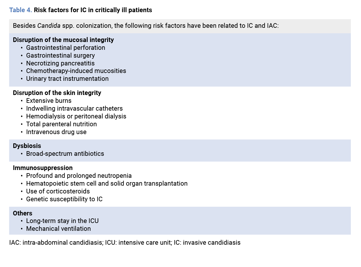 Different studies have also specifically addressed the risk factors for developing IAC. A retrospective case-control study, performed in 26 European ICUs from 2015 to 2016, included adult patients diagnosed with a microbiologically documented IAC (cases) and patients who had not developed IAC (controls) [3].
A total of 101 cases and 101 controls were included. C. albicans was the most common isolated species (58.4%), followed by C. glabrata (15.8%). Interestingly, concomitant blood cultures were only positive in 7 patients, and 16.8% of patients had, at least, 2 different Candida species identified. A multivariate analysis identified recurrent gastrointestinal perforation, anastomotic leakage, abdominal drain and prior prescription of antifungals or antibiotics drugs for 7 or more days as risk factors for IAC [3]. A different single-centre, retrospective case-control study which included 250 adult patients diagnosed with an intra-abdominal infection (125 cases and 125 controls), also reported that, besides upper gastrointestinal surgery, exposure to corticosteroids (prednisone > 20 mg equivalent for > 2 weeks), and mechanical ventilation were independent risk factors for developing IAC [70].
Based on these identified risk factors, several predictive scoring systems for IAC have been proposed. Dupont et al. built a scoring system based on 4 parameters: length of stay before surgery for > 48 hours, peri-operative cardiovascular failure, generalized peritonitis and upper gastrointestinal tract perforation [71]. This score had a high NPV and was especially useful for discarding the need for empirical antifungal treatment. Significantly, patients diagnosed with IAC based on a yeast-positive peritoneal fluid culture had higher severity scores and a threefold increase in mortality risk [71]. Finally, Li et al. suggested a scoring system based on C-reactive protein-to-albumin ratio, neutrophil-to-lymphocyte ratio, BDG positivity, and some clinical factors (sepsis, total parenteral nutrition, broad-spectrum antibiotic and SOFA score) [72]. This score was principally aimed at identifying patients with high risk of developing IC, and that could benefit from early empirical antifungal treatment.
4.2. How does empirical treatment impact on mortality or the development of antifungal resistance?
As previously mentioned, IAC is a severe disease associated with prolonged ICU and hospital stays, as well as an increased risk of mortality. Therefore, optimizing treatment is crucial to improving outcomes for patients diagnosed with IAC. A retrospective, multicentre, multinational study, conducted across 13 hospitals in 4 countries over a three-year period (2011-2013), addressed the risk factors associated with mortality in patients with IAC [5]. Adult patients admitted in surgical wards, ICUs and medical wards, such as internal medicine, hematology, or oncology were included. IAC was diagnosed according to the following criteria: Candida detection or growth from purulent or necrotic intra-abdominal specimens (including surgical, percutaneous aspiration and biliary samples, and/or biopsies obtained from intra-abdominal samples), Candida isolated from blood cultures in patients with secondary and tertiary peritonitis, and Candida growth from drainage tubes if placed less than 24 hours before the cultures were obtained. A total of 481 patients were included in the study. Overall, 252 (52.4%) and 131 (27.2%) patients were hospitalized in the surgical ward and the ICU, respectively. C. albicans (64%) and C. glabrata (16%) were the most commonly isolated yeasts. Echinocandins were the most frequently prescribed initial antifungal agent (63.8%). The 30-day mortality rate was 26.8%. In the multivariate analysis, several factors were independently associated with an increased risk of mortality, including age (OR 1.05), APACHE II score at diagnosis (OR 1.05), secondary peritonitis (OR 1.72), septic shock (OR 3.29), lack of source control (OR 3.35), and inadequate antifungal therapy (OR 1.81) [5]. Similar results were observed in a single-centre, retrospective study conducted in Pittsburgh, USA, which analysed 163 cases of IAC over two years [73]. The most common types of IAC were intra-abdominal abscesses (55%, 89/163) and secondary peritonitis (33%, 53/163). The 30-day mortality rate was 20% (32/163). Among all patients with IAC, younger age, the presence of an abscess and early source control were independently associated with survival. However, when focusing specifically on cases of secondary peritonitis or abscesses originating from gastrointestinal tract sources, early antifungal therapy was independently associated with survival (OR 0.3 for mortality).
Echinocandin resistance is an emerging clinical problem and is especially challenging in the cases of C. glabrata [74]. As previously reviewed, the abdominal cavity has been identified as a potential source of. C. glabrata drug-resistant isolates, and the emergence of resistance was mostly associated with insufficient antifungal exposure [36]. Additionally, a gastrointestinal colonization and systemic dissemination model for C. glabrata performed in mice demonstrated that treatment with echinocandins could lead to the development of resistant mutants within the gastrointestinal tract [75]. These mutant clones could afterwards disseminate. As such, antifungal stewardship and optimization of therapy, including empirical treatment, is mandatory.
Several studies have assessed the use of BDG for guiding antifungal treatment. A randomized, multicentre, controlled clinical trial conducted between 2016 and 2019 across 18 ICUs investigated the value of BDG in guiding early antifungal treatment in septic patients at high risk for IC [76]. Patients in the control group (n = 167) received targeted antifungal therapy driven by culture results, whereas those in the BDG group (n = 172) received antifungals if at least one of two consecutive BDG samples taken during the first two study days was ≥ 80 pg/mL. Although antifungal use was significantly higher in the BDG group compared to the control group (48.8% vs 12.0%), there were no differences in hospital or ICU length of stay, nor in 28-day mortality between the two groups. The authors concluded that this strategy could lead to unnecessary treatments and increased cost per patient, at least with the reported incidence of IC (14%) [76]. Nevertheless, other studies have shown that BDG may be useful for safely discontinuing empirical antifungal treatment [77].
An open-label clinical trial randomly assigned septic patients receiving empirical antifungal treatment for presumed Candida infection into two groups: one in which antifungal treatment was stopped if BDG was negative (cases) and another in which therapy continued based on clinical criteria (controls) [77]. A total of 53 patients were included in the BDG group, while 55 were in the control group. The number of complications during follow-up and the 30-day mortality rate were similar between both groups (28.3% [BDG group] vs 27.3% [control group], P = 0.92). However, patients in the BDG group had a significantly shorter duration of antifungal treatment (median [IQR]: 2 days [1-3] days vs 10 [6-13] days, P < 0.001). Additionally, there was also a notable decrease in antifungal treatment costs with echinocandins (708€ [185.6-1071.5] vs 1320€ [618.5-30,149.5], P = 0.07). The authors concluded that BDG could serve as a reliable antifungal stewardship tool in critically ill septic patients at risk of IC by reducing the duration of the empirical antifungal therapy [77]. Nonetheless, further studies are warranted.
4.3. What are the guideline recommendations on this entity? What is missing from these guidelines?
Most guidelines recommend echinocandins for the empirical treatment of Candida infections [78], while L-AmB should be prescribed if there is intolerance or resistance to echinocandins and azoles [7,79]. Specifical choice of antifungal therapy for IAC is commonly in line with recommendations for candidemia [80]. However, evidence comparing the different antifungals in IAC is missing, and there are concerns regarding echinocandin efficacy in this setting. Azoles, for susceptible Candida spp., and L-AmB, for azole-
resistant species, might be adequate alternatives.
Unfortunately, as previously reviewed, several unanswered questions remain concerning the optimization of the diagnosis and management of IC in the ICU, such as the identification of patients at risk, drug dosing and monitoring of treatment response [65]. The EPICO.4 was a Spanish high-level consensus document based on the Delphi methodology, involving 60 specialists from different hospitals. A series of recommendations were issued regarding the optimization of the management of non-neutropenic critically ill patients at risk of developing invasive candidiasis. Moreover, an easy-to-follow algorithm (the MAGICS algorithm) for the diagnosis and treatment of IC in critically ill patients was also established [81]. The EPICO.3 specifically addressed the diagnosis and management of post-surgical patients with complicated intra-abdominal infection and surgical patients with prolonged ICU stays. An easy-to-follow algorithm was also established for these patients [82].
4.4. Could the treatment be individualized?
IAC remain a challenging infection to manage in critically ill patients, with several areas of uncertainty. While guidelines have helped to standardize treatment approaches, they often lack specific recommendations for critically ill patients. Consequently, many experts now advocate for individualized treatment strategies based on the severity of infection, the site of infection, and the isolated pathogenic microorganism, with the goal of optimizing the antifungal drug exposure at the site of the infection [10,83]. L-AmB exhibits strong activity against Candida spp., has a very low risk of inducing antifungal resistance, and is effective against biofilm formation [10]. Given these advantages and the therapeutic challenges posed by IAC, it is reasonable to consider a potential role for L-AmB in specific clinical scenarios (Figure 1), including: i) Patients with deep-seated candidiasis or infections in sites where antifungal penetration and diffusion is limited (e.g., central nervous system, endocarditis, endophthalmitis, peritonitis), particularly when caused by echinocandin- or azole-resistant species such as C. krusei, C. glabrata, C. auris, or C. parapsilosis; ii) Patients with catheter-associated candidiasis (intravenous or peritoneal) where catheter removal is challenging (e.g., ECMO) or with intra-abdominal abscesses that cannot be surgically or percutaneously drained; iii) Patients with mixed fungal infections (e.g., yeast and filamentous fungi) [83]; iv) Patients previously treated with echinocandins or fluconazole; v) Patients showing worsening BDG levels despite empirical or targeted antifungal treatment and apparently adequate source control.
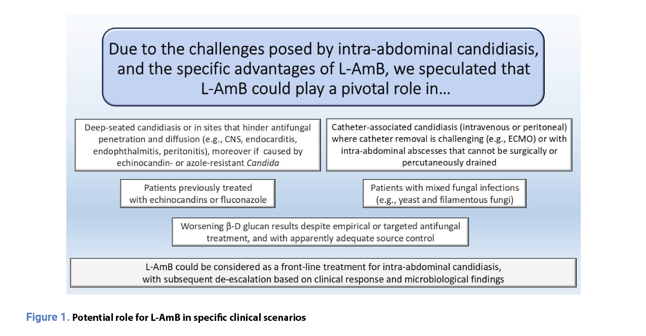 In fact, since early antifungal therapy is crucial to preventing biofilm formation and improving patient outcomes, some authors have suggested that L-AmB should be considered a front-line treatment for IAC, with subsequent de-escalation based on clinical response and microbiological findings [10]. Recently, a prospective, interventional phase 2 Italian study assessed the safety of pulsed high-dose L-AmB (5 mg/kg/day) in patients with suspected IAC managed with a BDG-guided strategy [84]. A total of 40 patients were enrolled. Following the microbiological tests at baseline (blood cultures, Gram stain and culture of intra-abdominal samples, and serum BDG determination), a loading dose of 5 mg/kg L-AmB was administered on day 1. On day 3, the decision to continue antifungal treatment dosage (standard dose 3 mg/kg/day) was based on the baseline BDG result. In the case of a negative baseline BDG result (< 80 pg/mL), antifungal therapy was discontinued. If the baseline BDG result was significantly positive (> 200 pg/mL) or IAC was confirmed by a culture result, the patient continued antifungal treatment for 7-14 days, as per the decision of the attending physician. In the case of a borderline positive BDG result (80-200 pg/mL), antifungal treatment was continued at the standard dose and was subsequently driven by BDG results on days 5, 7 and 14. Patients were followed up to 30 days after drug discontinuation. None of the patients with a negative baseline BDG result developed an IFI, whereas empirical antifungal therapy was stopped promptly. The authors concluded that a single high dose of L-AmB in critically ill patients with severe intra-abdominal disease was safe, and that when coupled with a BDG-guided strategy to rule out IC and/or IAC, it could lead to a reduction in antifungal exposure [84].
Based on these findings, a recent Italian consensus document promoted by the Multidisciplinary and Intersociety Italian Council for the Optimization of Antimicrobial Use, now recommends a pulse dose of L-AmB (5 mg/kg/day) as preemptive treatment in patients at high risk for IAC while BDG results are still pending [85].
5. Conclusions
IAC remains a significant challenge in critically ill patients, with high morbidity and mortality rates. Despite advancements in antifungal therapy and diagnostic methods, timely identification and appropriate treatment remain critical for improving patient outcomes. Standard microbiological diagnostics, including blood cultures, often lack sensitivity, leading to delays in treatment. Non-culture-based methods, such as β-D-glucan (BDG) and PCR, have shown promising in aiding early diagnosis and guiding antifungal therapy. However, their predictive value remains variable, necessitating further validation. The abdominal cavity has been identified as a hidden reservoir for antifungal resistance, particularly for echinocandins. Sequential collection of isolates for antifungal susceptibility testing seems essential. Critically ill ICU patients exhibit significant intra- and inter-individual variability in antifungal PK/PD. Obese and morbidly obese patients may require higher antifungal doses, except for L-AmB, whose clearance is unaffected by body weight. While fluconazole may need dose adjustments in patients undergoing RRT, echinocandins and L-AmB are not significantly impacted. ECMO can reduce blood levels of voriconazole and caspofungin but has minimal effect on L-AmB, which remains within the therapeutic range. These factors highlight the need for individualized dosing strategies in ICU patients. Empirical antifungal therapy is crucial for reducing mortality, particularly in patients with septic shock and high-risk features. However, unnecessary antifungal use can drive resistance and increase healthcare costs. Studies suggest that BDG-guided antifungal stewardship can safely reduce treatment duration without compromising outcomes. Echinocandins remain as the first-line antifungal agents for IAC, but their penetration into the peritoneal cavity is suboptimal, and resistance—especially in C. glabrata—is an emerging concern. L-AmB offers broad-spectrum activity, low resistance potential, and biofilm inhibition, making it a viable alternative in specific clinical settings. New antifungals, such as rezafungin and ibrexafungerp, offer improved pharmacokinetics and tissue penetration, but clinical experience remains limited. Future research should focus on optimizing antifungal selection, refining diagnostic algorithms, and enhancing treatment individualization based on patient-specific factors. Ultimately, a multidisciplinary approach combining rapid diagnostics, tailored antifungal therapy, and effective source control is essential for improving the management and prognosis of patients with IAC. Due to all these particularities, we suggest that specific guidelines focusing only on IAC are needed. Funding None to declare. Author contributions Conceptualization, all authors.; methodology, all authors; investigation, all authors; writing—original draft preparation, all authors; writing—review and editing, all authors; funding acquisition, all authors. All authors have read and agreed to the published version of the manuscript. Acknoweledgements Gilead supported the contribution of José Tiago Sequeira Lopes da Silva who helped to draft the manuscript Conflicts of Interest J.G. has received funds for participating in educational activities organised on behalf of Gilead, Pfizer, Mundipharma, and MSD; he has also received research funds from FIS, Gilead, F2G, Scynexis, Mundipharma, Cidara, and Shionogi outside the submitted work. P.P.-A. has received honoraria for talks on behalf of Merck Sharp and Dohme, Lilly, ViiV Healthcare and Gilead Science, and has participated in advisory boards for Gilead Science. E.M. has received funds for participating in educational activities organised on behalf of Gilead, Pfizer, Mundipharma, Viatris, Shionogi, and MSD. RZ has received funds for participating in educational activities organised on behalf of Gilead, Pfizer, and Mundipharma. The funders had no role in the design of the study; in the collection, analyses, or interpretation of data; in the writing of the manuscript; or in the decision to publish the results.References
- Soriano A, Honore PM, Puerta-Alcalde P, Garcia-Vidal C, Pagotto A, Goncalves-Bradley DC, et al. Invasive candidiasis: current clinical challenges and unmet needs in adult populations. J Antimicrob Chemother. 2023;78(7):1569-85. Available from: https://doi.org/10.1093/jac/dkad139
- Bongomin F, Gago S, Oladele RO, Denning DW. Global and multi-national prevalence of fungal diseases-estimate precision. J Fungi (Basel). 2017;3(4):57. Available from: https://doi.org/10.3390/jof3040057
- Bassetti M, Vena A, Giacobbe DR, Trucchi C, Ansaldi F, Antonelli M, et al. Risk factors for intra-abdominal candidiasis in intensive care units: results from EUCANDICU study. Infect Dis Ther. 2022;11(2):827-40. Available from: https://doi.org/10.1007/s40121-021-00585-6
- Montravers P, Dupont H, Gauzit R, Veber B, Auboyer C, Blin P, et al. Candida as a risk factor for mortality in peritonitis. Crit Care Med. 2006;34(3):646-52. Available from: https://doi.org/10.1097/01.CCM.0000201889.39443.D2
- Bassetti M, Righi E, Ansaldi F, Merelli M, Scarparo C, Antonelli M, et al. A multicenter multinational study of abdominal candidiasis: epidemiology, outcomes and predictors of mortality. Intensive Care Med. 2015; 41(9):1601-10. Available from: https://doi.org/10.1007/s00134-015-3866-2
- Montravers P, Mira JP, Gangneux JP, Leroy O, Lortholary O; AmarCand study. A multicentre study of antifungal strategies and outcome of Candida spp. peritonitis in intensive-care units. Clin Microbiol Infect. 2011;17(7): 1061-7. Available from: https://doi.org/10.1111/j.1469-0691.2010.03360.x
- Pappas PG, Kauffman CA, Andes DR, Clancy CJ, Marr KA, Ostrosky-Zeichner L, et al. Clinical practice guideline for the management of candidiasis: 2016 update by the Infectious Diseases Society of America. Clin Infect Dis. 2016;62(4):e1-50. Available from: https://doi.org/10.1093/cid/civ933
- Keighley C, Cooley L, Morris AJ, Ritchie D, Clark JE, Boan P, et al; Australasian Antifungal Guidelines Steering C. Consensus guidelines for the diagnosis and management of invasive candidiasis in haematology, oncology and intensive care settings, 2021. Intern Med J. 2021;51 Suppl 7:89-117. Available from: https://doi.org/10.1111/imj.15589
- Bassetti M, Giacobbe DR, Agvald-Ohman C, Akova M, Alastruey-Izquierdo A, Arikan-Akdagli S, et al. Invasive fungal diseases in adult patients in intensive care unit (FUNDICU): 2024 consensus definitions from ESGCIP, EFISG, ESICM, ECMM, MSGERC, ISAC, and ISHAM. Intensive Care Med. 2024;50(4):502-15. Available from: https://doi.org/10.1007/s00134-024-07341-7
- Maseda E, Martin-Loeches I, Zaragoza R, Peman J, Fortun J, Grau S, et al. Critical appraisal beyond clinical guidelines for intra-abdominal candidiasis. Crit Care. 2023;27(1):382. Available from: https://doi.org/10.1186/s13054-023-04673-6
- Dadar M, Tiwari R, Karthik K, Chakraborty S, Shahali Y, Dhama K. Candida albicans - biology, molecular characterization, pathogenicity, and advances in diagnosis and control - an update. Microb Pathog. 2018;117:128-38. Available from: https://doi.org/10.1016/j.micpath.2018.02.028
- Clancy CJ, Nguyen MH. Diagnosing invasive candidiasis. J Clin Microbiol. 2018;56(5):e01909-17. Available from: https://doi.org/10.1128/JCM.01909-17
- Leroy O, Gangneux JP, Montravers P, Mira JP, Gouin F, Sollet JP, et al. Epidemiology, management, and risk factors for death of invasive Candida infections in critical care: a multicenter, prospective, observational study in France (2005-2006). Crit Care Med. 2009;37(5):1612-8. Available from: https://doi.org/10.1097/CCM.0b013e31819efac0
- Fernandez J, Erstad BL, Petty W, Nix DE. Time to positive culture and identification for Candida bloodstream infections. Diagn Microbiol Infect Dis. 2009;64(4):402-7. Available from: https://doi.org/10.1016/j.diagmicrobio.2009.04.002
- Nunes CZ, Marra AR, Edmond MB, da Silva Victor E, Pereira CA. Time to blood culture positivity as a predictor of clinical outcome in patients with Candida albicans bloodstream infection. BMC Infect Dis. 2013;13:486. Available from: https://doi.org/10.1186/1471-2334-13-486
- Morrell M, Fraser VJ, Kollef MH. Delaying the empiric treatment of Candida bloodstream infection until positive blood culture results are obtained: a potential risk factor for hospital mortality. Antimicrob Agents Chemother. 2005;49(9):3640-5. Available from: https://doi.org/10.1128/AAC.49.9.3640-3645.2005
- Lagunes L, Rey-Perez A, Martin-Gomez MT, Vena A, de Egea V, Munoz P, et al. Association between source control and mortality in 258 patients with intra-abdominal candidiasis: a retrospective multi-centric analysis comparing intensive care versus surgical wards in Spain. Eur J Clin Microbiol Infect Dis. 2017;36(1):95-104. Available from: https://doi.org/10.1007/s10096-016-2775-9
- Alves J, Alonso-Tarres C, Rello J. How to identify invasive candidemia in ICU - a narrative review. Antibiotics (Basel). 2022;11(12):1804. Available from: https://doi.org/10.3390/antibiotics11121804
- Karageorgopoulos DE, Vouloumanou EK, Ntziora F, Michalopoulos A, Rafailidis PI, Falagas ME. Beta-D-glucan assay for the diagnosis of invasive fungal infections: a meta-analysis. Clin Infect Dis. 2011;52(6):750-70. Available from: https://doi.org/10.1093/cid/ciq206
- Onishi A, Sugiyama D, Kogata Y, Saegusa J, Sugimoto T, Kawano S, et al. Diagnostic accuracy of serum 1,3-beta-D-glucan for Pneumocystis jiroveci pneumonia, invasive candidiasis, and invasive aspergillosis: systematic review and meta-analysis. J Clin Microbiol. 2012;50(1):7-15. Available from: https://doi.org/10.1128/JCM.05267-11
- Lu Y, Chen YQ, Guo YL, Qin SM, Wu C, Wang K. Diagnosis of invasive fungal disease using serum (1→3)-beta-D-glucan: a bivariate meta-analysis. Intern Med. 2011;50(22):2783-91. Available from: https://doi.org/10.2169/internalmedicine.50.6175
- Fortun J, Buitrago MJ, Gioia F, Gomez-Garcia-de-la-Pedrosa E, Alvarez ME, Martin-Davila P, et al. Roles of the multiplex real-time PCR assay and beta-D-glucan in a high-risk population for intra-abdominal candidiasis (IAC). Med Mycol. 2020;58(6):789-96. Available from: https://doi.org/10.1093/mmy/myz123
- Dupont H, Malaquin S, Villeret L, Macq PY, Ammenouche N, Tinturier F, et al. Is ss-D-glucan relevant for the diagnosis and follow-up of intensive care patients with yeast- complicated intra-abdominal infection? J Fungi (Basel). 2022;8(5):487. Available from: https://doi.org/10.3390/jof8050487
- Novy E, Riviere J, Nguyen M, Arfeuille G, Louis G, Bouhemad B, et al. Combination of serum and peritoneal 1,3-beta-D-glucan can rule out intra-abdominal candidiasis in surgical critically ill patients: a multicenter prospective study. Crit Care. 2023;27(1):470. Available from: https://doi.org/10.1186/s13054-023-04761-7
- Tissot F, Lamoth F, Hauser PM, Orasch C, Fluckiger U, Siegemund M, et al; Fungal Infection Network of Switzerland. Beta-glucan antigenemia anticipates diagnosis of blood culture-negative intra-abdominal candidiasis. Am J Respir Crit Care Med. 2013;188(9):1100-9.Available from: https://doi.org/10.1164/rccm.201211-2069OC
- Leon C, Ruiz-Santana S, Saavedra P, Castro C, Ubeda A, Loza A, et al. Value of beta-D-glucan and Candida albicans germ tube antibody for discriminating between Candida colonization and invasive candidiasis in patients with severe abdominal conditions. Intensive Care Med. 2012;38(8):1315-25. Available from: https://doi.org/10.1007/s00134-012-2616-y
- Martinez-Jimenez MC, Muñoz P, Valerio M, Alonso R, Martos C, Guinea J, et al. Candida biomarkers in patients with candidaemia and bacteraemia. J Antimicrob Chemother. 2015;70(8):2354-61. Available from: https://doi.org/10.1093/jac/dkv090
- Xie M, Shao J, Wan Z, Yan T, Zhu S, Li S, et al. Detection of Candida DNA in peritoneal fluids by PCR assay optimizing the diagnosis and treatment for intra-abdominal candidiasis in high-risk ICU patients: a prospective cohort study. Front Microbiol. 2022;13:1070688. Available from: https://doi.org/10.3389/fmicb.2022.1070688
- Clancy CJ, Pappas PG, Vazquez J, Judson MA, Kontoyiannis DP, Thompson GR, et al. Detecting Infections Rapidly and Easily for Candidemia Trial, Part 2 (DIRECT2): a prospective, multicenter study of the T2Candida panel. Clin Infect Dis. 2018;66(11):1678-86. Available from: https://doi.org/10.1093/cid/cix1095
- Giannella M, Pankey GA, Pascale R, Miller VM, Miller LE, Seitz T. Antimicrobial and resource utilization with T2 magnetic resonance for rapid diagnosis of bloodstream infections: systematic review with meta-analysis of controlled studies. Expert Rev Med Devices. 2021;18(5):473-82. Available from: https://doi.org/10.1080/17434440.2021.1919508
- Honore PM, Redant S, Preseau T, Moorthamers S, Kaefer K, Barreto Gutierrez L, et al. T2MR can be used as a non-culture-based test together with biomarkers to improve detection of Candida in the bloodstream and reduce time delay in treating invasive candidiasis. Expert Rev Anti Infect Ther. 2022;20(3):327-9. Available from: https://doi.org/10.1080/14787210.2021.1964954
- Krifors A, Ullberg M, Castegren M, Petersson J, Sparrelid E, Hammarstrom H, et al. T2Candida assay in the diagnosis of intraabdominal candidiasis: a prospective multicenter study. J Fungi (Basel). 2022;8(1):832. Available from: https://doi.org/10.3390/jof8080832
- Lamoth F, Clancy CJ, Tissot F, Squires K, Eggimann P, Fluckiger U, et al. Performance of the T2Candida panel for the diagnosis of intra-abdominal candidiasis. Open Forum Infect Dis. 2020;7(3):ofaa075. Available from: https://doi.org/10.1093/ofid/ofaa075
- Diaz-Garcia J, Mesquida A, Machado M, Sanchez-Carrillo C, Munoz P, Escribano P, et al. Yeasts from blood cultures in the wake of the COVID-19 pandemic in a tertiary care hospital: shift in species epidemiology, steady low antifungal resistance and full in vitro ibrexafungerp activity. Med Mycol. 2023;61(7):myad072. Available from: https://doi.org/10.1093/mmy/myad072
- Diaz-Garcia J, Gomez A, Machado M, Alcala L, Reigadas E, Sanchez-Carrillo C, et al. Blood and intra-abdominal Candida spp. from a multicentre study conducted in Madrid using EUCAST: emergence of fluconazole resistance in Candida parapsilosis, low echinocandin resistance and absence of Candida auris. J Antimicrob Chemother. 2022;77(11):3102-9. Available from: https://doi.org/10.1093/jac/dkac288
- Diaz-Garcia J, Mesquida A, Gomez A, Machado M, Martin-Rabadan P, Alcala L, et al. Antifungal susceptibility testing identifies the abdominal cavity as a source of Candida glabrata-resistant isolates. Antimicrob Agents Chemother. 2021;65(12):e0124921. Available from: https://doi.org/10.1128/AAC.01249-21
- Diaz-Garcia J, Machado M, Alcala L, Reigadas E, Sanchez-Carrillo C, Perez-Ayala A, et al. Antifungal resistance in Candida spp. within the intra-abdominal cavity: study of resistance acquisition in patients with serial isolates. Clin Microbiol Infect. 2023;29(12):1604.e1-1604.e6. Available from: https://doi.org/10.1016/j.cmi.2023.08.021
- Shields RK, Nguyen MH, Press EG, Clancy CJ. Abdominal candidiasis is a hidden reservoir of echinocandin resistance. Antimicrob Agents Chemother. 2014;58(12):7601-5. Available from: https://doi.org/10.1128/AAC.04134-14
- Dzierba AL, Abrams D, Brodie D. Medicating patients during extracorporeal membrane oxygenation: the evidence is building. Crit Care. 2017;21(1):66. Available from: https://doi.org/10.1186/s13054-017-1644-y
- Jamal JA, Economou CJ, Lipman J, Roberts JA. Improving antibiotic dosing in special situations in the ICU: burns, renal replacement therapy and extracorporeal membrane oxygenation. Curr Opin Crit Care. 2012;18(5):460-71. Available from: https://doi.org/10.1097/MCC.0b013e32835685ad
- Sinnollareddy MG, Roberts JA, Lipman J, Akova M, Bassetti M, De Waele JJ, et al. Pharmacokinetic variability and exposures of fluconazole, anidulafungin, and caspofungin in intensive care unit patients: data from multinational Defining Antibiotic Levels in Intensive care unit (DALI) patients study. Crit Care. 2015;19(1):33. Available from: https://doi.org/10.1186/s13054-015-0758-3
- Van Daele R, Wauters J, Elkayal O, Dreesen E, Debaveye Y, Lagrou K, et al. Liposomal amphotericin B exposure in critically ill patients: a prospective pharmacokinetic study. Med Mycol. 2022;60(10):myac074. Available from: https://doi.org/10.1093/mmy/myac074
- NCD Risk Factor Collaboration (NCD-RisC). Trends in adult body-mass index in 200 countries from 1975 to 2014: a pooled analysis of 1698 population-based measurement studies with 19.2 million participants. Lancet. 2016;387(10026):1377-96. Available from: https://doi.org/10.1016/S0140-6736(16)30054-X
- Alobaid AS, Wallis SC, Jarrett P, Starr T, Stuart J, Lassig-Smith M, et al. Effect of obesity on the population pharmacokinetics of fluconazole in critically ill patients. Antimicrob Agents Chemother. 2016;60(11):6550-7. Available from: https://doi.org/10.1128/AAC.01088-16
- Maseda E, Grau S, Luque S, Castillo-Mafla MP, Suarez-de-la-Rica A, Montero-Feijoo A, et al. Population pharmacokinetics/pharmacodynamics of micafungin against Candida species in obese, critically ill, and morbidly obese critically ill patients. Crit Care. 2018;22(1):94. Available from: https://doi.org/10.1186/s13054-018-2019-8
- Wasmann RE, Ter Heine R, van Dongen EP, Burger DM, Lempers VJ, Knibbe CA, et al. Pharmacokinetics of anidulafungin in obese and normal-weight adults. Antimicrob Agents Chemother. 2018;62(7):e00063-18. Available from: https://doi.org/10.1128/AAC.00063-18
- Ferriols-Lisart R, Aguilar G, Perez-Pitarch A, Puig J, Ezquer-Garin C, Alos M. Plasma concentrations of caspofungin in a critically ill patient with morbid obesity. Crit Care. 2017;21(1):200. Available from: https://doi.org/10.1186/s13054-017-1774-2
- Wasmann RE, Smit C, van Dongen EPH, Wiezer RMJ, Adler-Moore J, de Beer YM, et al. Fixed dosing of liposomal amphotericin B in morbidly obese individuals. Clin Infect Dis. 2020;70(10):2213-5. Available from: https://doi.org/10.1093/cid/ciz885
- Yagasaki K, Gando S, Matsuda N, Kameue T, Ishitani T, Hirano T, et al. Pharmacokinetics and the most suitable dosing regimen of fluconazole in critically ill patients receiving continuous hemodiafiltration. Intensive Care Med. 2003;29(10):1844-8. Available from: https://doi.org/10.1007/s00134-003-1980-z
- Leitner JM, Meyer B, Fuhrmann V, Saria K, Zuba C, Jager W, et al. Multiple-dose pharmacokinetics of anidulafungin during continuous venovenous haemofiltration. J Antimicrob Chemother. 2011;66(4):880-4. Available from: https://doi.org/10.1093/jac/dkq545
- Aguilar G, Azanza JR, Carbonell JA, Ferrando C, Badenes R, Parra MA, et al. Anidulafungin dosing in critically ill patients with continuous venovenous haemodiafiltration. J Antimicrob Chemother. 2014;69(6):1620-3. Available from: https://doi.org/10.1093/jac/dkt542
- Maseda E, Grau S, Villagran MJ, Hernandez-Gancedo C, Lopez-Tofino A, Roberts JA, et al. Micafungin pharmacokinetic/pharmacodynamic adequacy for the treatment of invasive candidiasis in critically ill patients on continuous venovenous haemofiltration. J Antimicrob Chemother. 2014;69(6):1624-32. Available from: https://doi.org/10.1093/jac/dku013
- Weiler S, Seger C, Pfisterer H, Stienecke E, Stippler F, Welte R, et al. Pharmacokinetics of caspofungin in critically ill patients on continuous renal replacement therapy. Antimicrob Agents Chemother. 2013;57(8):4053-7. Available from: https://doi.org/10.1128/AAC.00335-13
- Obata Y, Takazono T, Tashiro M, Ota Y, Wakamura T, Takahashi A, et al. The clinical usage of liposomal amphotericin B in patients receiving renal replacement therapy in Japan: a nationwide observational study. Clin Exp Nephrol. 2021;25(3):279-87. Available from: https://doi.org/10.1007/s10157-020-01989-3
- Shekar K, Roberts JA, McDonald CI, Ghassabian S, Anstey C, Wallis SC, et al. Protein-bound drugs are prone to sequestration in the extracorporeal membrane oxygenation circuit: results from an ex vivo study. Crit Care. 2015;19(1):164. Available from: https://doi.org/10.1186/s13054-015-0891-z
- Ruiz S, Papy E, Da Silva D, Nataf P, Massias L, Wolff M, et al. Potential voriconazole and caspofungin sequestration during extracorporeal membrane oxygenation. Intensive Care Med. 2009;35(1):183-4. Available from: https://doi.org/10.1007/s00134-008-1269-3
- Gioia F, Gomez-Lopez A, Alvarez ME, Gomez-Garcia de la Pedrosa E, Martin-Davila P, Cuenca-Estrella M, et al. Pharmacokinetics of echinocandins in suspected Candida peritonitis: a potential risk for resistance. Int J Infect Dis. 2020;101:24-8. Available from: https://doi.org/10.1016/j.ijid.2020.09.019
- Pea F, Righi E, Cojutti P, Carnelutti A, Baccarani U, Soardo G, et al. Intra-abdominal penetration and pharmacodynamic exposure to fluconazole in three liver transplant patients with deep-seated candidiasis. J Antimicrob Chemother. 2014;69(9):2585-6. Available from: https://doi.org/10.1093/jac/dku169
- Tortora F, Dei Giudici L, Simeoli R, Chiusolo F, Cairoli S, Bernaschi P, et al. Therapeutic drug monitoring of amphotericin B in plasma and peritoneal fluid of pediatric patients after liver transplantation: a case series. Antibiotics (Basel). 2022;11(5):640. Available from: https://doi.org/10.3390/antibiotics11050640
- Novy E, Roger C, Roberts JA, Cotta MO. Pharmacokinetic and pharmacodynamic considerations for antifungal therapy optimisation in the treatment of intra-abdominal candidiasis. Crit Care. 2023;27(1):449. Available from: https://doi.org/10.1186/s13054-023-04742-w
- Lee A, Prideaux B, Zimmerman M, Carter C, Barat S, Angulo D, et al. Penetration of ibrexafungerp (formerly SCY-078) at the site of infection in an intra-abdominal candidiasis mouse model. Antimicrob Agents Chemother. 2020;64(3):e02268-19. Available from: https://doi.org/10.1128/AAC.02268-19
- Lal S, Kumar-Singh V, Agarwal S. Candida isolation from peritoneal fluid: its role in the outcome of patients with perforation peritonitis. J Emerg Pract Trauma. 2022;8(1):49-54. Available from: https://doi.org/10.34172/jept.2021.20
- Felk A, Kretschmar M, Albrecht A, Schaller M, Beinhauer S, Nichterlein T, et al. Candida albicans hyphal formation and the expression of the Efg1-regulated proteinases Sap4 to Sap6 are required for the invasion of parenchymal organs. Infect Immun. 2002;70(7):3689-700. Available from: https://doi.org/10.1128/IAI.70.7.3689-3700.2002
- Lass-Florl C, Kanj SS, Govender NP, Thompson GR 3rd, Ostrosky-Zeichner L, Govrins MA. Invasive candidiasis. Nat Rev Dis Primers. 2024;10(1):20. Available from: https://doi.org/10.1038/s41572-024-00503-3
- Logan C, Martin-Loeches I, Bicanic T. Invasive candidiasis in critical care: challenges and future directions. Intensive Care Med. 2020;46(11):2001-14. Available from: https://doi.org/10.1007/s00134-020-06240-x
- Del Palacio A, Villar J, Alhambra A. Epidemiology of invasive candidiasis in pediatric and adult populations. Rev Iberoam Micol. 2009;26(1):2-7. Available from: https://doi.org/10.1016/S1130-1406(09)70002-6
- Thomas-Ruddel DO, Schlattmann P, Pletz M, Kurzai O, Bloos F. Risk factors for invasive Candida infection in critically ill patients: a systematic review and meta-analysis. Chest. 2022;161(2):345-55. Available from: https://doi.org/10.1016/j.chest.2021.08.081
- Alenazy H, Alghamdi A, Pinto R, Daneman N. Candida colonization as a predictor of invasive candidiasis in non-neutropenic ICU patients with sepsis: a systematic review and meta-analysis. Int J Infect Dis. 2021;102:357-62. Available from: https://doi.org/10.1016/j.ijid.2020.10.092
- Park JY, Yang KM, Kwak JY, Jung YT. Risk factors for invasive candidiasis in critically ill patients who underwent emergency gastrointestinal surgery for complicated intra-abdominal infection. Surg Infect (Larchmt). 2024;25(4):307-14. Available from: https://doi.org/10.1089/sur.2023.333
- Habighorst K, Sanders JM, Hennessy SA, Goff K, Wan B, Johns M. Identification of risk factors for intra-abdominal candidiasis. Surg Infect (Larchmt). 2023;24(10):910-5. Available from: https://doi.org/10.1089/sur.2023.149
- Dupont H, Guilbart M, Ntouba A, Perquin M, Petiot S, Regimbeau JM, et al. Can yeast isolation be predicted in complicated secondary non-postoperative intra-abdominal infections? Crit Care. 2015;19(1):60. Available from: https://doi.org/10.1186/s13054-015-0790-3
- Li W, Chen G, Lin F, Yang H, Cui Y, Lu R, et al. A scoring system based on novel biomarkers and clinical risk factors to predict invasive candidiasis in immunocompetent critically ill patients. Front Microbiol. 2023;14:1097574. Available from: https://doi.org/10.3389/fmicb.2023.1097574
- Vergidis P, Clancy CJ, Shields RK, Park SY, Wildfeuer BN, Simmons RL, et al. Intra-abdominal candidiasis: the importance of early source control and antifungal treatment. PLoS One. 2016;11(4):e0153247. Available from: https://doi.org/10.1371/journal.pone.0153247
- Arendrup MC, Perlin DS. Echinocandin resistance: an emerging clinical problem? Curr Opin Infect Dis. 2014;27(6):484-92. Available from: https://doi.org/10.1097/QCO.0000000000000111
- Healey KR, Nagasaki Y, Zimmerman M, Kordalewska M, Park S, Zhao Y, et al. The gastrointestinal tract is a major source of echinocandin drug resistance in a murine model of Candida glabrata colonization and systemic dissemination. Antimicrob Agents Chemother. 2017;61(12):e01412-17. Available from: https://doi.org/10.1128/AAC.01412-17
- Bloos F, Held J, Kluge S, Simon P, Kogelmann K, de Heer G, et al. (1→3)-beta-D-glucan-guided antifungal therapy in adults with sepsis: the CandiSep randomized clinical trial. Intensive Care Med. 2022;48(7):865-75. Available from: https://doi.org/10.1007/s00134-022-06733-x
- De Pascale G, Posteraro B, D’Arrigo S, Spinazzola G, Gaspari R, Bello G, et al. (1,3)-beta-D-glucan-based empirical antifungal interruption in suspected invasive candidiasis: a randomized trial. Crit Care. 2020;24(1):550. Available from: https://doi.org/10.1186/s13054-020-03265-y
- Bassetti M, Peghin M, Timsit JF. The current treatment landscape: candidiasis. J Antimicrob Chemother. 2016;71 Suppl 2:ii13-22. Available from: https://doi.org/10.1093/jac/dkw392
- Martin-Loeches I, Antonelli M, Cuenca-Estrella M, Dimopoulos G, Einav S, De Waele JJ, et al. ESICM/ESCMID task force on practical management of invasive candidiasis in critically ill patients. Intensive Care Med. 2019;45(6):789-805. Available from: https://doi.org/10.1007/s00134-019-05599-w
- Cornely OA, Sprute R, Bassetti M, Chen SC, Groll AH, Kurzai O, et al. Global guideline for the diagnosis and management of candidiasis: an initiative of the ECMM in cooperation with ISHAM and ASM. Lancet Infect Dis. 2025. Available from: https://doi.org/10.1016/S1473-3099(24)00749-7
- Zaragoza R, Ferrer R, Llinares P, Maseda E, Rodriguez A, Grau S, et al. EPICO 4.0. Total quality in the management of invasive candidiasis in critically ill patients by analysing the integrated process. Rev Iberoam Micol. 2017;34(3):143-57. Available from: https://doi.org/10.1016/j.riam.2017.03.008
- Maseda E, Rodriguez AH, Aguilar G, Peman J, Zaragoza R, Ferrer R, et al. EPICO 3.0. Recommendations on invasive candidiasis in patients with complicated intra-abdominal infection and surgical patients with ICU extended stay. Rev Iberoam Micol. 2016;33(4):196-205. Available from: https://doi.org/10.1016/j.riam.2016.02.003
- Zaragoza R, Maseda E, Peman J. Individualized antifungal therapy in critically ill patients with invasive fungal infection. Rev Iberoam Micol. 2021;38(2):68-74. Available from: https://doi.org/10.1016/j.riam.2021.04.006
- Rinaldi M, Bartoletti M, Bonazzetti C, Caroccia N, Gatti M, Tazza B, et al. Tolerability of pulsed high-dose L-AmB as pre-emptive therapy in patients at high risk for intra-abdominal candidiasis: a phase 2 study (LAMBDA study). Int J Antimicrob Agents. 2023;62(6):106998. Available from: https://doi.org/10.1016/j.ijantimicag.2023.106998
- Sartelli M, Tascini C, Coccolini F, Dellai F, Ansaloni L, Antonelli M, et al. Management of intra-abdominal infections: recommendations by the Italian council for the optimization of antimicrobial use. World J Emerg Surg. 2024;19(1):23. Available from: https://doi.org/10.1186/s13017-024-00551-w
Explore
Articles
Volumes
About the journal
Most read
Recomendaciones para la prevención y el tratamiento de las infecciones respiratorias en adultos mayores institucionalizados
Revista Española de Quimioterapia
Acute infectious gastroenteritis in childhood: the role of rapid multiplex molecular syndromic panels in diagnosis and clinical management
Revista Española de Quimioterapia
Características epidemiológicas de pacientes con aislamientos de Enterobacterales productores de carbapenemasas-NDM en un hospital de alta complejidad
Revista Española de Quimioterapia
Review on the management of intra-abdominal candidiasis
Revista Española de Quimioterapia
Neoscytalidium dimidiatum: a rare locally acquired deep cutaneous infection in a renal transplant recipient
Revista Española de Quimioterapia