Letter
April 2025, Volume 38, nº 2
9 min
Systemic absorption of liposomal inhaled amikacin in the treatment of resistant Mycobacterium avium: beyond expectations
Belén Sánchez-Pascual*1, Juan Emilio Losa-García2, Sira Sanz-Márquez1, Irene Salvador-Llana1, Ana Vegas-Serrano2, Montserrat Pérez-Encinas1
1Servicio de Farmacia, Hospital Universitario Fundación Alcorcón, Alcorcón, Madrid, España
2Servicio de Infecciosas, Hospital Universitario Fundación Alcorcón, Alcorcón, Madrid, España
http://www.doi.org/10.37201/req/115.2024
Receipt Date:
02/12/2024
Acceptance Date:
16/02/2025
Publication Date:
25/02/2025
How to cite
Sánchez Pascual B, Losa García JE, Sanz Márquez S, Salvador Llana I, Vegas Serrano A, Pérez Encinas M. Systemic absorption of liposomal inhaled amikacin in the treatment of resistant Mycobacterium avium: beyond expectations. Rev Esp Quimioter. 2025 Feb 26;38(2):148-151. doi: 10.37201/req/115.2024
Metrics
637
Views
55
Downloads
Share
Share
Dear Editor,
M. avium causes common non-tuberculous mycobacterial (NTM) lung infections, with rising incidence due to limited knowledge and therapeutic options [1-3]. It typically affects patients with pre-existing pulmonary or immune-related conditions [1,4,5]. However, less frequently, it can also occur in immunocompetent individuals without underlying pathologies.
M. avium’s treatment is highly toxic, prolonged (lasting up to 12 months post culture negativization) [1,2] and involves complex drug regimens [3]. M. avium employs immune evasion mechanisms and forms biofilms in the airways, to ensure pulmonary persistence [4]. Moreover, intrinsic resistance and acquired resistance to other antimicrobials, due to a high genomic mutation rate, further complicate treatment [4]. Current guidelines advise determining strain susceptibility before selecting the regimen [6].
Treatment involves a three-drug regimen comprising: macrolides, if strain is susceptible [2,3], as they improve response rates compared to first-line anti-tuberculosis drugs [1] (azithromycin, preferred over clarithromycin due to better tolerance, fewer interactions, and once-daily dosing), ethambutol, and rifampicin [1].
Parenteral aminoglycosides are not typically recommended for mild to moderate cases susceptible to macrolides. However, they are advised if disease is cavitary or advanced, with severe bronchiectasis, or if there is macrolide resistance [2,3], where culture conversion rates are lower (15-36%) [4]. Amikacin penetrates M. avium biofilms and macrophages [3], strengthening the pharmacological regimen [7]. However, its low lung penetration rate [4,8] requires high doses to achieve effective lung concentrations, increasing toxicity and limiting its use [7].
To enhance efficacy and tolerability, inhaled liposomal amikacin (ILA) has been developed, offering a nebulized formulation with similar composition to lung surfactant [3,4]. This route allows for higher lung concentrations and lower blood concentrations, enhancing efficacy and reducing systemic toxicity. Liposomes act as nanocarriers, allowing controlled drug release and aiding access to macrophages and biofilms [2,4].
In CONVERT study, adding ILA to conventional therapy (CT), demonstrated increased sputum culture conversion (SCC) rates compared to CT alone: 29% vs 8.9% (ODSS ratio 4.22, 95% CI 2.08-8.57, p<0.0001) [4,9]. SCC, defined as three consecutive negative sputum cultures after six months of treatment, was the primary endpoint. TR02-112 study also showed a significant culture negativization (32% vs 9%) compared with placebo [8]. Consequently, current guidelines recommend adding ILA if positive sputum persists after 6 months of CT [1,3,8].
Common adverse effects (AE) of ILA include respiratory issues: cough, dysphonia, and hemoptysis. Ototoxicity was higher compared to CT [9], possibly due to drug accumulation, although overall accumulation appears insignificant [4].
ILA kinetics follows a tricompartmental model [4]. In CONVERT and TR02-112 trials, involving 53 patients, maximum concentrations (Cmax) was < 2 mg/L initially and after 6 months [8]. Cmax increased at 6 months, but by less than 10%, suggesting minimal accumulation [8]. Systemic exposure was relatively low, median Cmax <2 mg/L (0.47-6.87 mg/L), notably lower than those achieved through parenteral administration (35-77 mg/L) [3,8].
A 57-year-old non-smoking woman with a hiatal hernia, visited the Emergency Department of her reference hospital in October 2019 with sudden hemoptoic cough, linked to bleeding from the hiatal hernia. Due to persistent episodes, a fibrobronchoscopy was performed, showing bronchiectasis. Cultures were positive for M. avium susceptible to clarithromycin.
On 27/01/2020, she started the treatment recommended by guidelines: azithromycin 500 mg (3 days/week), ethambutol 1400 mg (3 days/week), and rifampicin 600 mg (3 days/week) for 12 months.
A follow-up scan in March 2021 showed no interstitial involvement but confirmed bronchiectasis. In May, she received the SARS-CoV-2 vaccine. Two weeks later, hemoptysis returned, with new infection signs on a July scan. Serratia was isolated, leading to treatment with ciprofloxacin and sulfamethoxazole. Additionally, M. avium, resistant to linezolid, clarithromycin, and moxifloxacin but susceptible to amikacin, was also isolated.
In November 2021, she looked for a second opinion at our Infectious Diseases Department. Clinical assessment indicated recurrent pulmonary disease due to mycobacteria. In January 2022, the diagnosis of pulmonary infection by M. avium resistant to clarithromycin and susceptible to amikacin was confirmed.
On 04/04/2022, she began treatment with parenteral amikacin (PA) 1 g daily, ILA 590 mg daily (sourced from Spanish Agency of Medicines), ethambutol (2 tablets/day, 400 mg each), rifampicin (2 tablets/day, 300 mg each), and moxifloxacin (1 tablet/day, 400 mg). This regimen was chosen due to macrolide resistance and prior 12-month treatment with subsequent relapse. Given the high failure rate in NTM pulmonary infections treatment, particularly with resistant forms, parenteral aminoglycosides were added to enhance treatment’s effectiveness.
Baseline assessment by Ophthalmology and Otorhinolaryngology found normal audiometry and visual acuity. Bi-monthly toxicity monitoring was established. Pharmacokinetic monitoring of amikacin was planned by the Pharmacy Service, aiming for minimum levels <1 mg/L to avoid toxicity and peaks >45 mg/L for efficacy assurance. The first pharmacokinetic assessment showed a minimum of 1.55 mg/L and a peak of 43.36 mg/L Adjustments using the Abbottbase-Pharmacokinetic-System (PKS)® were attempted, but accuracy was limited due to unadjusted analytical data and patient characteristics.
Elevated minimum suggested lower-than-expected drug elimination for a woman of her characteristics compared to the reference population. Initially, it was suspected that patient might have lower-than-expected renal elimination based on her usual creatinine levels (0.6-0.8 mg/dl). It was assumed that glomerular filtration (GFR) might be impaired for reasons other than creatinine levels.
The treatment plan was changed to 1 g/48 hours of PA, while maintaining daily ILA. Pharmacokinetic adjustments improved levels (lowered minimum, raised peak). However, subsequent analyses again detected minimums above 1 mg/L and insufficient peaks, prompting additional dose adjustments (Figure 1). Levels never matched PKS® estimations. No nephrotoxic factors were found, suggesting filtration aligned with creatinine levels.
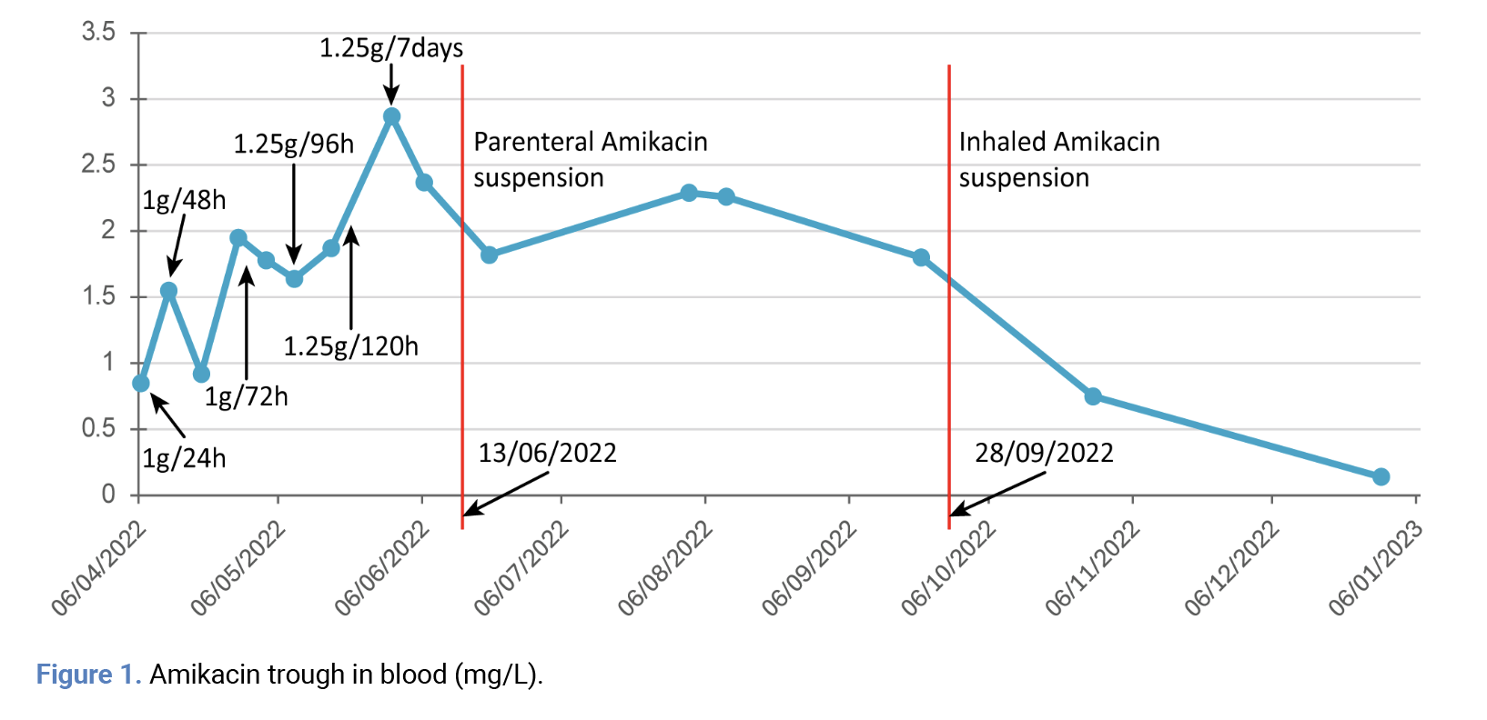 The possibility that ILA might experience higher-than-expected absorption, not accounted for by PKS®, causing accumulation was then considered. It was decided to end PA administration. Last PA dose was on 13/06/2022. A week later, a minimum and a mid-concentration (after 12 hours) were measured, yielding 1.82 and 2.82 mg/L, respectively.
Positive cultures in May and June 2022 led to a treatment update, exploring potential macrolide resensitization with consultations to the National Microbiology Center. Given the positive response, moxifloxacin was switched to clarithromycin due to arthralgia.
New regimen, started on 25/08/2022: ILA daily, ethambutol (2 tablets/day, 400 mg each), rifampicin (2 tablets/day, 300 mg each), and azithromycin (1 tablet/day, 250 mg).
Cultures were negative from 05/07/2023 onwards. After PA’s discontinuation, amikacin minimum levels remained high. After five months of treatment, ILA was suspended due to suspected ototoxicity identified during a follow-up by the Otorhinolaryngology Department. Amikacin continued to be detected in blood up to 3 months post-ILA suspension.
During treatment, the patient experienced dysphonia due to acute laryngitis and pharyngolaryngeal reflux, which resolved rest and a visit to a phoniatrist. Subsequently, she developed oral candidiasis, treated with nystatin, and vaginal candidiasis, resolved with topical treatment and clotrimazole ovules. A cough persisted post-treatment, GFR remained stable, and neurosensory hearing loss persisted after ILA suspension.
ILA, a novel NTM pulmonary infection treatment, offers higher target levels and improves macrophage penetration due to its unique administration route and liposomal structure. Technical sheet (TS) doesn’t reflect significant alveolar absorption, and there’s little information regarding its monitoring.
In our patient, treatment with both PA and ILA were initiated considering she had been previously treated and relapsed (with gained macrolide resistance). SCC in strains resistant to macrolides are lower than those for susceptible strains, so the controlled risk of combining both presentations was assumed to increase treatment’s likelihood of success.
To manage amikacin, pharmacokinetic analysis was performed, initially not considering that ILA could influence, as its absorption was deemed negligible according to TS. However, analytical determinations of amikacin minimum levels were unexpectedly elevated for a woman of her characteristics and PA doses. This raised the possibility that patient might have worse elimination than reflected by her creatinine levels, which was ruled out as she was not exposed to any nephrotoxic agents, had a good overall condition with no clinical indicators of GFR deterioration, and that the PKS® adjustments estimated concentrations that were not reflected in real life.
It was then assumed that systemic absorption of ILA exceeded expectations, leading to accumulation. PA was discontinued and amikacin blood levels were determined 1 week after suspension. The most significant finding was that while TS reflected an average peak of 1.81 mg/L, our patient presented a minimum level of 1.81 mg/L one week after PA suspension. Previous treatment with PA could falsify minimum levels by elevating them but considering that serum half-life of PA is slightly over 2 hours [10] and PA treatment had ended one week earlier, it should have been completely eliminated, and the minimum would only correspond to the contribution of ILA.
Amikacin persisted in bloodstream for up to 3 months after ILA cessation, suggesting its accumulation [4]. Monitoring amikacin’s blood peak post-ILA administration lacks efficacy assessment since ILA targets its therapeutic site directly. Evaluating blood concentrations for efficacy elsewhere is counterproductive (although sputum monitoring would be interesting). Few pharmacokinetic analyses regarding blood concentrations of ILA focus on maximum concentrations, ranging from 0.47-6.87 mg/L [3,8], without assessing minimum, essential for toxicity control, probably because with such low peaks, minimal concentrations are expected to be negligible, making monitoring unnecessary.
In our experience, the disparity between minimum and maximum concentrations post-ILA administration is not as pronounced as in intravenous administration, with minimum concentrations nearing peaks. Monitoring minimum amikacin blood levels after ILA administration aids in systemic toxicity surveillance, enhancing AE prevention and management.
Funding
None to declare.
Conflict of interest
Authors declare no conflict of interest.
Author contributions
B.S.P.; writing—original draft preparation, investigation, J.E.L.G.; investigation, S.S.M.; investigation, writing—review and editing, I.S.L.; investigation, writing—review and editing, A.V.S.; investigation, M.P.E.; writing—review and editing, investigation. All authors have read and agreed to the published version of the manuscript.
The possibility that ILA might experience higher-than-expected absorption, not accounted for by PKS®, causing accumulation was then considered. It was decided to end PA administration. Last PA dose was on 13/06/2022. A week later, a minimum and a mid-concentration (after 12 hours) were measured, yielding 1.82 and 2.82 mg/L, respectively.
Positive cultures in May and June 2022 led to a treatment update, exploring potential macrolide resensitization with consultations to the National Microbiology Center. Given the positive response, moxifloxacin was switched to clarithromycin due to arthralgia.
New regimen, started on 25/08/2022: ILA daily, ethambutol (2 tablets/day, 400 mg each), rifampicin (2 tablets/day, 300 mg each), and azithromycin (1 tablet/day, 250 mg).
Cultures were negative from 05/07/2023 onwards. After PA’s discontinuation, amikacin minimum levels remained high. After five months of treatment, ILA was suspended due to suspected ototoxicity identified during a follow-up by the Otorhinolaryngology Department. Amikacin continued to be detected in blood up to 3 months post-ILA suspension.
During treatment, the patient experienced dysphonia due to acute laryngitis and pharyngolaryngeal reflux, which resolved rest and a visit to a phoniatrist. Subsequently, she developed oral candidiasis, treated with nystatin, and vaginal candidiasis, resolved with topical treatment and clotrimazole ovules. A cough persisted post-treatment, GFR remained stable, and neurosensory hearing loss persisted after ILA suspension.
ILA, a novel NTM pulmonary infection treatment, offers higher target levels and improves macrophage penetration due to its unique administration route and liposomal structure. Technical sheet (TS) doesn’t reflect significant alveolar absorption, and there’s little information regarding its monitoring.
In our patient, treatment with both PA and ILA were initiated considering she had been previously treated and relapsed (with gained macrolide resistance). SCC in strains resistant to macrolides are lower than those for susceptible strains, so the controlled risk of combining both presentations was assumed to increase treatment’s likelihood of success.
To manage amikacin, pharmacokinetic analysis was performed, initially not considering that ILA could influence, as its absorption was deemed negligible according to TS. However, analytical determinations of amikacin minimum levels were unexpectedly elevated for a woman of her characteristics and PA doses. This raised the possibility that patient might have worse elimination than reflected by her creatinine levels, which was ruled out as she was not exposed to any nephrotoxic agents, had a good overall condition with no clinical indicators of GFR deterioration, and that the PKS® adjustments estimated concentrations that were not reflected in real life.
It was then assumed that systemic absorption of ILA exceeded expectations, leading to accumulation. PA was discontinued and amikacin blood levels were determined 1 week after suspension. The most significant finding was that while TS reflected an average peak of 1.81 mg/L, our patient presented a minimum level of 1.81 mg/L one week after PA suspension. Previous treatment with PA could falsify minimum levels by elevating them but considering that serum half-life of PA is slightly over 2 hours [10] and PA treatment had ended one week earlier, it should have been completely eliminated, and the minimum would only correspond to the contribution of ILA.
Amikacin persisted in bloodstream for up to 3 months after ILA cessation, suggesting its accumulation [4]. Monitoring amikacin’s blood peak post-ILA administration lacks efficacy assessment since ILA targets its therapeutic site directly. Evaluating blood concentrations for efficacy elsewhere is counterproductive (although sputum monitoring would be interesting). Few pharmacokinetic analyses regarding blood concentrations of ILA focus on maximum concentrations, ranging from 0.47-6.87 mg/L [3,8], without assessing minimum, essential for toxicity control, probably because with such low peaks, minimal concentrations are expected to be negligible, making monitoring unnecessary.
In our experience, the disparity between minimum and maximum concentrations post-ILA administration is not as pronounced as in intravenous administration, with minimum concentrations nearing peaks. Monitoring minimum amikacin blood levels after ILA administration aids in systemic toxicity surveillance, enhancing AE prevention and management.
Funding
None to declare.
Conflict of interest
Authors declare no conflict of interest.
Author contributions
B.S.P.; writing—original draft preparation, investigation, J.E.L.G.; investigation, S.S.M.; investigation, writing—review and editing, I.S.L.; investigation, writing—review and editing, A.V.S.; investigation, M.P.E.; writing—review and editing, investigation. All authors have read and agreed to the published version of the manuscript.
References
- Esteban J, Navas E. Tratamiento de las infecciones producidas por micobacterias no tuberculosas. Enferm Infecc Microbiol Clin (Engl Ed). 2018 Nov;36(9):586-592. doi: 10.1016/j.eimc.2017.10.008.
- Shirley M. Amikacin Liposome Inhalation Suspension: A Review in Mycobacterium avium Complex Lung Disease. Drugs. 2019 Mar 15;79(5):555–62. doi: 10.1007/s40265-019-01095-z
- Adjemian J, Daniel-Wayman S, Ricotta E, Prevots DR. Epidemiology of Nontuberculous Mycobacteriosis. Semin Resp Crit Care. 2018 Jun;39(03):325–35. doi: 10.1055/s-0038-1651491
- Chalmers JD, van Ingen J, van der Laan R, Herrmann JL. Liposomal drug delivery to manage nontuberculous mycobacterial pulmonary disease and other chronic lung infections. Eur Respir Rev. 2021 Jul 20;30(161):210010. doi: 10.1183/16000617.0010-2021.
- Adjemian J, Daniel-Wayman S, Ricotta E, Prevots DR. Epidemiology of Nontuberculous Mycobacteriosis. Semin Respir Crit Care Med. 2018 Jun;39(03):325–35. doi: 10.1055/s-0038-1651491
- WHO consolidated guidelines on tuberculosis. Module 4: treatment - drug-resistant tuberculosis treatment, 2022 update [Internet]. www.who.int. Available from: https://www.who.int/publications/i/item/9789240063129
- Jaleh Varshosaz, Ghaffari S, Mirshojaei SF, Jafarian A, Atyabi F, Kobarfard F, Azarmi S. Biodistribution of Amikacin Solid Lipid Nanoparticles after Pulmonary Delivery. BioMed Res Int. 2013 Jan 1;2013:1–8. doi: 10.1155/2013/136859
- Rubino CM, Onufrak NJ, van Ingen J, Griffith DE, Bhavnani SM, Yuen DW, et al. Population Pharmacokinetic Evaluation of Amikacin Liposome Inhalation Suspension in Patients with Treatment-Refractory Nontuberculous Mycobacterial Lung Disease. Eur J Drug Metab Ph. 2021 Feb 17;46(2):277–87. doi: 10.1007/s13318-020-00669-7
- Griffith DE, Eagle G, Thomson R, Aksamit TR, Hasegawa N, Morimoto K, et al. Amikacin Liposome Inhalation Suspension for Treatment-Refractory Lung Disease Caused by Mycobacterium avium Complex (CONVERT). A Prospective, Open-Label, Randomized Study. Am J Respir Crit Care Med. 2018 Dec 15;198(12):1559–69. doi: 10.1164/rccm.201807-1318OC.
- FICHA TECNICA AMIKACINA NORMON 500 MG/2 ML SOLUCION INYECTABLE Y PARA PERFUSIÓN EFG [Internet]. cima.aemps.es. Available from: https://cima.aemps.es/cima/dochtml/ft/57012/FichaTecnica_57012.html.
Explore
Articles
Volumes
About the journal
Most read
Recomendaciones para la prevención y el tratamiento de las infecciones respiratorias en adultos mayores institucionalizados
Revista Española de Quimioterapia
Acute infectious gastroenteritis in childhood: the role of rapid multiplex molecular syndromic panels in diagnosis and clinical management
Revista Española de Quimioterapia
Características epidemiológicas de pacientes con aislamientos de Enterobacterales productores de carbapenemasas-NDM en un hospital de alta complejidad
Revista Española de Quimioterapia
Review on the management of intra-abdominal candidiasis
Revista Española de Quimioterapia
Neoscytalidium dimidiatum: a rare locally acquired deep cutaneous infection in a renal transplant recipient
Revista Española de Quimioterapia