Letter
April 2026, Volume 39, nº 2
7 min
Visceral leishmaniasis in a multiple sclerosis patient: a rare complication of fingolimod-induced lymphopenia
Alba García-Alabarce1, Elena Cornejo-Calvo2  , Adolfo de Salazar-González3,4,5, Emilio Guirao-Arrabal4,6*
1Internal Medicine Unit, Hospital Santa Ana, Motril, Spain
2Hematology Unit, Hospital Universitario Clínico San Cecilio, Granada, Spain
3Microbiology Unit, Hospital Universitario Clínico San Cecilio, Granada, Spain
4Instituto de Investigación Biosanitaria ibs.GRANADA, Granada, Spain
5CIBER de Enfermedades Infecciosas (CIBERINFEC), Instituto de Salud Carlos III, Madrid, Spain
6Infectious Diseases Unit, Hospital Universitario Clínico San Cecilio, Granada, Spain
*Correspondence:
Emilio Guirao-Arrabal. emilio.guirao@gmail.com
Receipt Date:
09/11/2025
Acceptance Date:
19/02/2026
Publication Date:
08/03/2026
How to cite
García-Alabarce A, Cornejo-Calvo E, de Salazar-González A, Guirao-Arrabal E. Visceral leishmaniasis in a multiple sclerosis patient: a rare complication of fingolimod-induced lymphopenia. Rev Esp Quimioter 2026; 39(2):148-150. Available from: http://www.doi.org/10.37201/req/122.2025
Metrics
910
Views
129
Downloads
Share
Share
Sir,
We present the case of a 56-year-old male resident of a rural area with a personal medical history that includes hypertension, chronic kidney disease, smoking-related interstitial lung disease, and relapsing-remitting multiple sclerosis (MS) that was treated with the immunomodulatory drug fingolimod.
The patient presented to the emergency department of our hospital with a complaint of fever reaching up to 39°C over the preceding seven days, accompanied by profuse sweating, without chills. The patient also exhibited symptoms of moderate fatigue but no loss of appetite or weight.
A physical examination was conducted, with the following findings: the patient’s temperature was 37.5°C, blood pressure 130/88 mmHg, heart rate 107 beats per minute (bpm), and arterial oxygen saturation levels were 95% while breathing room air. A subsequent abdominal examination revealed hepatosplenomegaly, measured at 5 cm below the costal margin for both organs, which did not cause discomfort.
The initial blood test revealed stable renal failure, with creatinine levels of 1.8 mg/dL, and marked pronounced pancytopenia, with a haemoglobin concentration of 9.8 g/dL, 2,830 leukocytes/μL, and a total lymphocyte count of 330/μL, as well as 73,000 platelets/μL. The C-reactive protein level was 149 mg/L, and the procalcitonin level was 0.98 ng/mL. A chest X-ray showed no abnormalities. Initial microbiological studies yielded negative results, including polymerase chain reaction (PCR) for respiratory viruses in nasal swabs (RSV, SARS-CoV-2, and influenza), urine antigen tests for Streptococcus pneumoniae and Legionella pneumophila, and blood and urine cultures.
The patient was subsequently transferred to our Infectious Diseases Unit without antimicrobial treatment pending definitive diagnosis, and fingolimod was discontinued. Antinuclear antibodies, extractable nuclear antigens, and anti-dsDNA were all reported as negative. Immunoglobulins were found to be within normal limits. Furthermore, serological analysis for HIV, hepatitis B, hepatitis C, EBV, CMV, parvovirus B19, Rickettsia conorii and Coxiella burnetii (IgG and IgM) returned negative results. Conversely, serological analysis for Leishmania spp. revealed a positive result (Leishmania VirClia® IgG+IgM, Vircell).
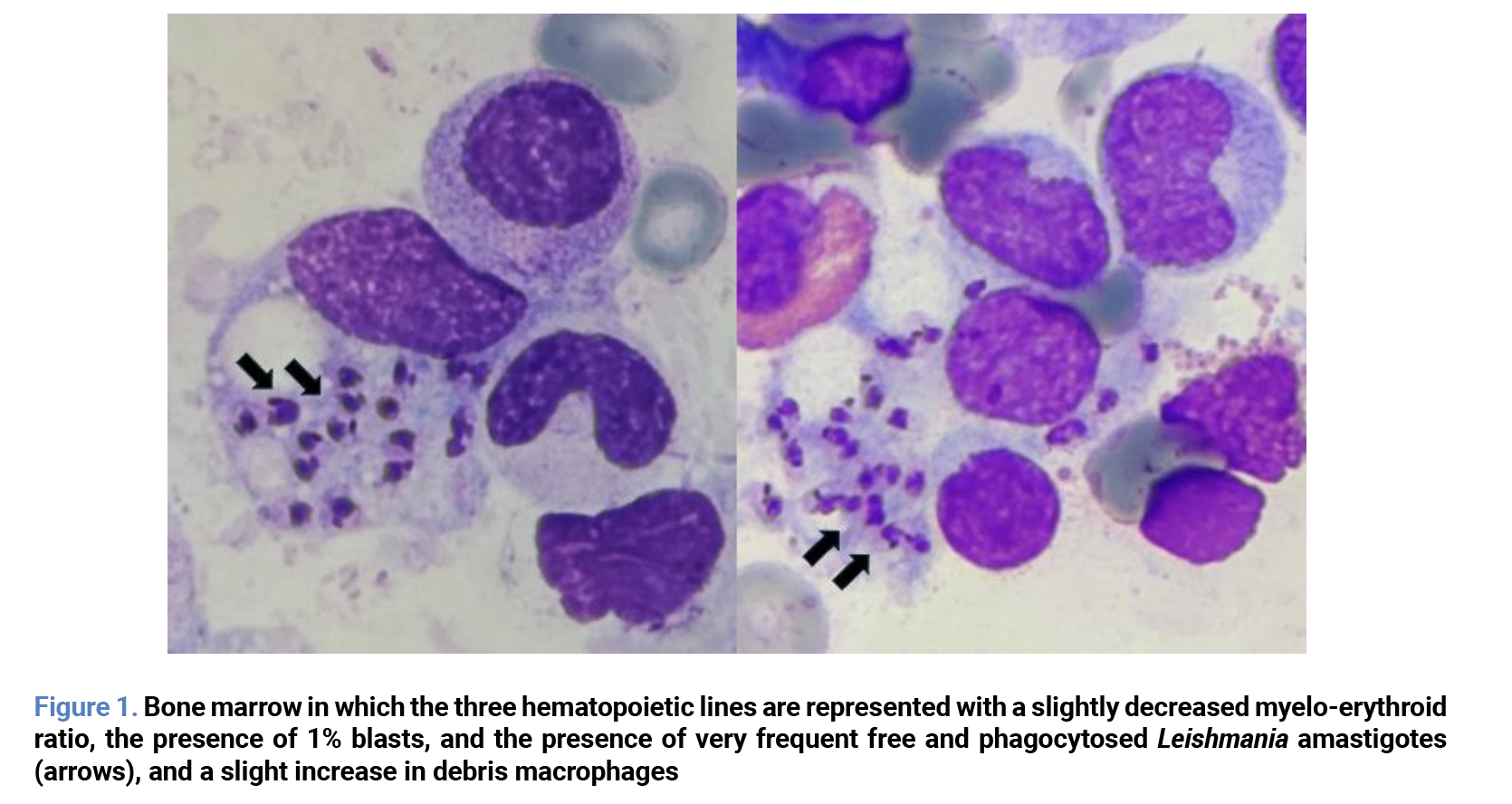
A bone marrow aspiration was then performed, and direct microscopic visualization revealed images consistent with the presence of Leishmania amastigotes (Figure 1). Confirmation was made using a real-time PCR technique (VIASURE Leishmania Real-Time PCR Detection Kit, Certest) on a bone marrow sample. Consequently, a definitive diagnosis of visceral leishmaniasis (VL) was established.
Liposomal amphotericin B at 5 mg/kg/day IV was administered on days 0, 5, and 10, which was well tolerated, and a favourable evolution of the blood count was observed. Subsequently, a booster dose was administered on day +30. One month after fingolimod discontinuation and VL therapy was started, the total lymphocyte count was 1,120/µl.
The causative agent of VL in the Mediterranean countries is the protozoan Leishmania infantum, which is transmitted to humans by the bite of a phlebotomine sandfly. Leishmaniasis is a widely distributed disease with higher incidences in Latin America, the Middle East, and Asia, and it is also endemic in the Mediterranean countries, including Spain.
In Spain, the predominant phlebotomine species is Phlebotomus perniciosus, which is distributed throughout the country and exhibits seasonal activity between May and November. The transmission of the disease is also subject to the presence of reservoirs, such as canines and small lagomorphs, including hares. Initial inoculation has been demonstrated to result in the manifestation of cutaneous forms, which subsequently persist as chronic latent infections.
In the province of Granada (Spain), a study revealed a 7.9% seropositivity for Leishmania spp. in donor sera [1]. While most people will never develop the disease, in situations of cellular immunosuppression, infection can reactivate, causing VL, which presents with fever, general symptoms, hepatosplenomegaly and pancytopenia.
In the year 2024, 473 cases of leishmaniasis were reported in Spain, 50.4% of which were classified as VL, with a mortality rate of 2.7%. It is noteworthy that this condition is likely underreported [2].
Immunosuppression is thus a major risk factor for the development of VL. During the 1990s and the early years of the 21st century, HIV was the prevailing factor associated with VL. In the contemporary era, individuals living with HIV no longer face a high risk of latent infections reactivating as a result of the high efficacy of modern antiretroviral therapies. At present, the cohort of patients demonstrating a high risk of reactivation of latent infections, such as VL, has been expanded to encompass other groups. These include chronic diseases requiring immunosuppressive therapies, solid organ transplantation, haematopoietic stem cell transplantation, and malignant haematological diseases [3].
One example of immunosuppressive treatment is MS, which has evolved significantly in recent years thanks to new disease-modifying therapies (DMT). The first drug in this group to be formulated orally was fingolimod. This drug inhibits sphingosine-1-phosphate receptors, causing sequestration of naïve and central memory T lymphocytes (CCR7 positive) in lymph nodes, leading to selective lymphopenia. To assess the risk to which patients on fingolimod therapy are exposed, we recommend monitoring lymphocyte subpopulations in follow-up blood tests. Overall, the risk of infection is no higher in patients treated with fingolimod, except in cases of severe or very severe lymphopenia (under 500 or under 200 cells/μL) [4].
Our patient was treated with a particular regimen of liposomal amphotericin B based on our initial clinical experience reported 20 years ago. This regimen consisted of four doses of 5 mg/kg (a total dose of 20 mg/kg) [5]. The WHO recommends a total dose of 18–21 mg/kg for Leishmania infantum regions, administered at a daily dose of 3–5 mg/kg for 3–6 days [6]. Although there are no clinical trials evaluating the preferred regimen for L. infantum, tolerance of liposomal amphotericin B appears excellent. Relapses appear to be associated with HIV infection and age rather than the dose or regimen used [7].
Regarding the risk of VL in MS patients under DMT, there are very few case reports [8,9]. Given the significant seroprevalence of latent leishmaniasis in our area and the risk of lymphopenia associated with fingolimod therapy, we aim to raise attention to this infection and its potential consequences if left undiagnosed and untreated. If VL is diagnosed in a patient taking fingolimod, in addition to initiating appropriate treatment for VL, we recommend switching to a different drug that does not pose such a risk.
Funding
None to declare.
Author contributions
Writing – original draft, A.G.-A.; Investigation, E.C.-C. and A.S.-G.; Writing – review & editing, E.G.-A.; Literature review, E.G.-A. All authors reviewed the successive versions of the manuscript.
Conflicts of interest
No commercial, financial, or personal conflicts of interest were identified in relation to the research, authorship, and/or publication of this article.
Ethics statement
Written informed consent for publication was obtained from the patient in accordance with the Declaration of Helsinki.
AI statement
ChatGPT (OpenAI, version 5.2) was used solely to assist with English language editing. The authors reviewed and approved all outputs generated by the tool and take full responsibility for the content of the manuscript.
References
- Aliaga L, Ceballos J, Sampedro A, Cobo F, López-Nevot MÁ, Merino-Espinosa G, et al. Asymptomatic Leishmania infection in blood donors from the Southern of Spain. Infection. 2019;47(5):739–47. Available from: https://doi.org/10.1007/s15010-019-01297-3
- CIBER de Epidemiología y Salud Pública; Centro Nacional de Epidemiología; Instituto de Salud Carlos III. Informe epidemiológico sobre la situación de la leishmaniasis en España, 2024. Madrid: ISCIII; 2025 Nov.
- Van Griensven J, Carrillo E, López-Vélez R, Lynen L, Moreno J. Leishmaniasis in immunosuppressed individuals. Clin Microbiol Infect. 2014;20(4):286–99. Available from: https://doi.org/10.1111/1469-0691.12556
- Francis G, Kappos L, O’Connor P, Collins W, Tang D, Mercier F, et al. Temporal profile of lymphocyte counts and relationship with infections with fingolimod therapy. Mult Scler J. 2014;20(4):471–80. Available from: https://doi.org/10.1177/1352458513500551
- Canora Lebrato J, Troncoso García E, Escobar T, Hernández Quero J. Treatment of visceral leishmaniasis in HIV patients with a new regimen of liposomal amphotericin. Med Clin. 2001;116(10):395. Available from: https://doi.org/10.1016/S0025-7753(01)71839-8
- van Griensven J, Diro E. Visceral Leishmaniasis: Recent Advances in Diagnostics and Treatment Regimens. Infect Dis Clin North Am 2019;33(1):79–99. Available from: https://doi.org/10.1016/j.idc.2018.10.005
- Masi F Di, Ursini T, Iannece MD, Chianura L, Baldasso F, Foti G, et al. Five-year retrospective Italian multicenter study of visceral leishmaniasis treatment. Antimicrob Agents Chemother. 2014;58(1):414–8. Available from: https://doi.org/10.1128/AAC.00840-13
- Guerra-Amor A, Lopez-Gonzalez A, Bosch-Nicolau P, Rodriguez-Acevedo B, Zabalza A, Sulleiro E, et al. Case Report: Leishmaniasis in a 33-Year-Old Man with Multiple Sclerosis. Am J Trop Med Hyg. 2022;107(2):339–41. Available from: https://doi.org/10.4269/ajtmh.22-0117
- Williams E, Isles NS, Seemann T, Kilpatrick T, Grigg A, Leroi M, et al. Case report: Confirmation by metagenomic sequencing of visceral leishmaniasis in an immunosuppressed returned traveler. Am J Trop Med Hyg. 2020;103(5):1930–3. Available from: https://doi.org/10.4269/ajtmh.19-0841
Explore
Articles
Volumes
About the journal
Most read
Recomendaciones para la prevención y el tratamiento de las infecciones respiratorias en adultos mayores institucionalizados
Revista Española de Quimioterapia
Acute infectious gastroenteritis in childhood: the role of rapid multiplex molecular syndromic panels in diagnosis and clinical management
Revista Española de Quimioterapia
Características epidemiológicas de pacientes con aislamientos de Enterobacterales productores de carbapenemasas-NDM en un hospital de alta complejidad
Revista Española de Quimioterapia
Review on the management of intra-abdominal candidiasis
Revista Española de Quimioterapia
Neoscytalidium dimidiatum: a rare locally acquired deep cutaneous infection in a renal transplant recipient
Revista Española de Quimioterapia